 Movie
Movie Controller

[English] 日本語
 Yorodumi
- PDB-1jup: Crystal structure of the multidrug binding transcriptional repres... -
+ Open data

- Basic information
| Entry | Database: PDB / ID: 1jup | ||||||
|---|---|---|---|---|---|---|---|
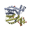
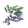
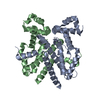
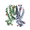
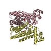
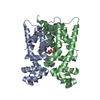
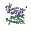
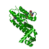
| Title | Crystal structure of the multidrug binding transcriptional repressor QacR bound to malachite green | ||||||
 Components Components | HYPOTHETICAL TRANSCRIPTIONAL REGULATOR IN QACA 5'REGION | ||||||
 Keywords Keywords | TRANSCRIPTION / multidrug recognition / malachite green / QacR / multidrug binding protein | ||||||
| Function / homology |  Function and homology information Function and homology informationDNA-binding transcription factor activity / negative regulation of DNA-templated transcription / DNA binding Similarity search - Function | ||||||
| Biological species |   Staphylococcus aureus (bacteria) Staphylococcus aureus (bacteria) | ||||||
| Method |  X-RAY DIFFRACTION / MOLECULAR REPLACEMENT / Resolution: 2.95 Å X-RAY DIFFRACTION / MOLECULAR REPLACEMENT / Resolution: 2.95 Å | ||||||
 Authors Authors | Schumacher, M.A. / Miller, M.C. / Grkovic, S. / Brown, M.H. / Skurray, R.A. / Brennan, R.G. | ||||||
 Citation Citation | Journal: Science / Year: 2001 Title: Structural mechanisms of QacR induction and multidrug recognition. Authors: Schumacher, M.A. / Miller, M.C. / Grkovic, S. / Brown, M.H. / Skurray, R.A. / Brennan, R.G. | ||||||
| History |
|
- Structure visualization
| Structure viewer | Molecule: MolmilJmol/JSmol |
|---|
- Downloads & links
-Download
| PDBx/mmCIF format | 1jup.cif.gz | 164.3 KB | Display | PDBx/mmCIF format |
|---|---|---|---|---|
| PDB format | pdb1jup.ent.gz | 132.6 KB | Display | PDB format |
| PDBx/mmJSON format | 1jup.json.gz | Tree view | PDBx/mmJSON format | |
| Others |  Other downloads Other downloads |
-Validation report
| Arichive directory | https://data.pdbj.org/pub/pdb/validation_reports/ju/1jupftp://data.pdbj.org/pub/pdb/validation_reports/ju/1jup | HTTPS FTP |
|---|
-Related structure data
| Related structure data |  1jt6C  1jtxC 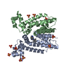 1jtyC 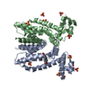 1jumC 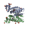 1jusSC C: citing same article ( S: Starting model for refinement |
|---|---|
| Similar structure data |


-Links
 PDBj
PDBj- Assembly
| Deposited unit | 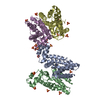
| ||||||||
|---|---|---|---|---|---|---|---|---|---|
| 1 | 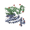
| ||||||||
| 2 | 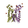
| ||||||||
| Unit cell |
| ||||||||
| Details | QacR is a dimer and there are two QacR dimers in the ASU one bound with the determined drug binding stoichiometry of one drug per dimer |
-Components
| #1: Protein | Mass: 22983.023 Da / Num. of mol.: 4 / Mutation: C72A, C141S Source method: isolated from a genetically manipulated source Source: (gene. exp.) Staphylococcus aureus (bacteria) / Plasmid: pSK5210 / Production host: #2: Chemical | ChemComp-SO4 /   Mass: 96.063 Da / Num. of mol.: 22 / Source method: obtained synthetically / Formula: SO4 Mass: 96.063 Da / Num. of mol.: 22 / Source method: obtained synthetically / Formula: SO4#3: Chemical | ChemComp-MGR / | 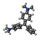  Mass: 329.458 Da / Num. of mol.: 1 / Source method: obtained synthetically / Formula: C23H25N2 / Comment: Antimicrobial*YM Mass: 329.458 Da / Num. of mol.: 1 / Source method: obtained synthetically / Formula: C23H25N2 / Comment: Antimicrobial*YM#4: Water | ChemComp-HOH / |  Mass: 18.015 Da / Num. of mol.: 7 / Source method: isolated from a natural source / Formula: H2O Mass: 18.015 Da / Num. of mol.: 7 / Source method: isolated from a natural source / Formula: H2O |
|---|
-Experimental details
-Experiment
| Experiment | Method: X-RAY DIFFRACTION / Number of used crystals: 1 |
|---|
- Sample preparation
| Crystal | Density Matthews: 3.97 Å3/Da / Density % sol: 69.04 % |
|---|---|
| Crystal grow | Temperature: 298 K / Method: vapor diffusion, hanging drop / pH: 7.5 Details: Ammonium sulphate, pH 7.5, VAPOR DIFFUSION, HANGING DROP, temperature 298K |
-Data collection
| Diffraction | Mean temperature: 298 K |
|---|---|
| Diffraction source | Source: ROTATING ANODE / Type: RIGAKU RU300 / Wavelength: 1.5418 Å |
| Detector | Type: RIGAKU RAXIS IV / Detector: IMAGE PLATE / Date: Nov 23, 2000 / Details: yale mirrors |
| Radiation | Monochromator: monochromator / Protocol: SINGLE WAVELENGTH / Monochromatic (M) / Laue (L): M / Scattering type: x-ray |
| Radiation wavelength | Wavelength: 1.5418 Å / Relative weight: 1 |
| Reflection | Resolution: 2.95→64.6 Å / Num. all: 26023 / Num. obs: 26023 / % possible obs: 86.8 % / Observed criterion σ(F): 0 / Observed criterion σ(I): 0 / Redundancy: 4 % / Biso Wilson estimate: 47.3 Å2 / Rmerge(I) obs: 0.089 / Rsym value: 0.089 / Net I/σ(I): 11 |
| Reflection shell | Resolution: 2.95→3.1 Å / Mean I/σ(I) obs: 2.4 / Rsym value: 0.278 / % possible all: 78 |
- Processing
| Software |
| ||||||||||||||||||||||||||||||||||||
|---|---|---|---|---|---|---|---|---|---|---|---|---|---|---|---|---|---|---|---|---|---|---|---|---|---|---|---|---|---|---|---|---|---|---|---|---|---|
| Refinement | Method to determine structure: MOLECULAR REPLACEMENT Starting model: PDB ENTRY 1JUS Resolution: 2.95→64.6 Å / Rfactor Rfree error: 0.006 / Data cutoff high absF: 1038053.03 / Data cutoff low absF: 0 / Isotropic thermal model: RESTRAINED / Cross valid method: THROUGHOUT / σ(F): 0 / σ(I): 0 / Stereochemistry target values: Engh and huber
| ||||||||||||||||||||||||||||||||||||
| Solvent computation | Solvent model: FLAT MODEL / Bsol: 43.9022 Å2 / ksol: 0.331763 e/Å3 | ||||||||||||||||||||||||||||||||||||
| Displacement parameters | Biso mean: 65 Å2
| ||||||||||||||||||||||||||||||||||||
| Refine analyze |
| ||||||||||||||||||||||||||||||||||||
| Refinement step | Cycle: LAST / Resolution: 2.95→64.6 Å
| ||||||||||||||||||||||||||||||||||||
| Refine LS restraints |
| ||||||||||||||||||||||||||||||||||||
| LS refinement shell | Resolution: 2.95→3.13 Å / Rfactor Rfree error: 0.025 / Total num. of bins used: 6
| ||||||||||||||||||||||||||||||||||||
| Xplor file |
|
