2-(3-amino-3-carboxypropyl)histidine synthase activity / oxidoreductase activity, acting on iron-sulfur proteins as donors / tRNA wobble base 5-methoxycarbonylmethyl-2-thiouridinylation / Synthesis of diphthamide-EEF2 / protein histidyl modification to diphthamide / tRNA wobble uridine modification / iron chaperone activity / ferrous iron binding / ubiquitin protein ligase activity / ubiquitin-dependent protein catabolic process ...2-(3-amino-3-carboxypropyl)histidine synthase activity / oxidoreductase activity, acting on iron-sulfur proteins as donors / tRNA wobble base 5-methoxycarbonylmethyl-2-thiouridinylation / Synthesis of diphthamide-EEF2 / protein histidyl modification to diphthamide / tRNA wobble uridine modification / iron chaperone activity / ferrous iron binding / ubiquitin protein ligase activity / ubiquitin-dependent protein catabolic process / protein ubiquitination / iron ion binding / zinc ion binding / nucleus / cytoplasm / cytosol Similarity search - Function
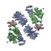
A: PROTEIN ATS1, DIPHTHAMIDE BIOSYNTHESIS PROTEIN 3 B: PROTEIN ATS1, DIPHTHAMIDE BIOSYNTHESIS PROTEIN 3 C: PROTEIN ATS1, DIPHTHAMIDE BIOSYNTHESIS PROTEIN 3 E: PROTEIN ATS1, DIPHTHAMIDE BIOSYNTHESIS PROTEIN 3 G: PROTEIN ATS1, DIPHTHAMIDE BIOSYNTHESIS PROTEIN 3 H: PROTEIN ATS1, DIPHTHAMIDE BIOSYNTHESIS PROTEIN 3 hetero molecules
Mass: 96.063 Da / Num. of mol.: 5 / Source method: obtained synthetically / Formula: SO4
Sequence details
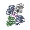
FUSION PROTEIN OF KTI13 (UNIPROT ID P31386, RESIDUES A1- A333) AND KTI11 (UNIPROT ID Q3E840, ...FUSION PROTEIN OF KTI13 (UNIPROT ID P31386, RESIDUES A1- A333) AND KTI11 (UNIPROT ID Q3E840, RESIDUES A344-A417) USING A GSGSGSGSGS LINKER (RESIDUES A334-A343), LINKER ORDERED FUSION PROTEIN OF KTI13 (UNIPROT ID P31386, RESIDUES G1- G333) AND KTI11 (UNIPROT ID Q3E840, RESIDUES G344-G417) USING A GSGSGSGSGS LINKER (RESIDUES G334-G343), LINKER ORDERED FUSION PROTEIN OF KTI13 (UNIPROT ID P31386, RESIDUES B1- B333) AND KTI11 (UNIPROT ID Q3E840, RESIDUES H344-H417) USING A GSGSGSGSGS LINKER (RESIDUES B334,B335,H343), LINKER PARTIALLY DISORDERED FUSION PROTEIN OF KTI13 (UNIPROT ID P31386, RESIDUES E1- E333) AND KTI11 (UNIPROT ID Q3E840, RESIDUES C344-C417) USING A GSGSGSGSGS LINKER (RESIDUES E334,C343), LINKER PARTIALLY DISORDERED
-
Experimental details
-
Experiment
Experiment
Method: X-RAY DIFFRACTION / Number of used crystals: 1
-
Sample preparation
Crystal
Density Matthews: 2.43 Å3/Da / Density % sol: 49.34 % / Description: NONE
Crystal grow
Details: 100 MM TRIS PH 8.4, 2 M LI2SO4
-
Data collection
Diffraction
Mean temperature: 100 K
Diffraction source
Source: SYNCHROTRON / Site: PETRA III, EMBL c/o DESY / Beamline: P14 (MX2) / Wavelength: 0.97626
In the structure databanks used in Yorodumi, some data are registered as the other names, "COVID-19 virus" and "2019-nCoV". Here are the details of the virus and the list of structure data.
Jan 31, 2019. EMDB accession codes are about to change! (news from PDBe EMDB page)
EMDB accession codes are about to change! (news from PDBe EMDB page)
The allocation of 4 digits for EMDB accession codes will soon come to an end. Whilst these codes will remain in use, new EMDB accession codes will include an additional digit and will expand incrementally as the available range of codes is exhausted. The current 4-digit format prefixed with “EMD-” (i.e. EMD-XXXX) will advance to a 5-digit format (i.e. EMD-XXXXX), and so on. It is currently estimated that the 4-digit codes will be depleted around Spring 2019, at which point the 5-digit format will come into force.
The EM Navigator/Yorodumi systems omit the EMD- prefix.
Related info.:Q: What is EMD? / ID/Accession-code notation in Yorodumi/EM Navigator
Yorodumi is a browser for structure data from EMDB, PDB, SASBDB, etc.
This page is also the successor to EM Navigator detail page, and also detail information page/front-end page for Omokage search.
The word "yorodu" (or yorozu) is an old Japanese word meaning "ten thousand". "mi" (miru) is to see.
Related info.:EMDB / PDB / SASBDB / Comparison of 3 databanks / Yorodumi Search / Aug 31, 2016. New EM Navigator & Yorodumi / Yorodumi Papers / Jmol/JSmol / Function and homology information / Changes in new EM Navigator and Yorodumi
 Movie
Movie Controller


 Open data
 Basic information
 Components
 Keywords
 Function and homology information
 X-RAY DIFFRACTION /
  Authors
 Citation
 Structure visualization
 Downloads & links
 Other downloads

 PDBj
PDBj
 Assembly
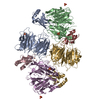


 Mass: 55.845 Da / Num. of mol.: 4 / Source method: obtained synthetically / Formula: Fe
 Mass: 96.063 Da / Num. of mol.: 5 / Source method: obtained synthetically / Formula: SO4
 Sample preparation
 / Beamline: P14 (MX2) / Wavelength: 0.97626
  Processing