 Movie
Movie Controller


+ Open data

- Basic information
| Entry | Database: PDB / ID: 4d1n | ||||||
|---|---|---|---|---|---|---|---|
| Title | Structure of human nNOS heme domain with L-Arg bound | ||||||
 Components Components | NITRIC OXIDE SYNTHASE, BRAIN | ||||||
 Keywords Keywords | OXIDOREDUCTASE | ||||||
| Function / homology |  Function and homology information Function and homology informationpositive regulation of membrane repolarization during ventricular cardiac muscle cell action potential / negative regulation of calcium ion transport into cytosol / Nitric oxide stimulates guanylate cyclase / myoblast fusion / ROS and RNS production in phagocytes / tetrahydrobiopterin binding / arginine binding / regulation of calcium ion transmembrane transport via high voltage-gated calcium channel / regulation of cardiac muscle contraction by calcium ion signaling / synaptic signaling by nitric oxide ...positive regulation of membrane repolarization during ventricular cardiac muscle cell action potential / negative regulation of calcium ion transport into cytosol / Nitric oxide stimulates guanylate cyclase / myoblast fusion / ROS and RNS production in phagocytes / tetrahydrobiopterin binding / arginine binding / regulation of calcium ion transmembrane transport via high voltage-gated calcium channel / regulation of cardiac muscle contraction by calcium ion signaling / synaptic signaling by nitric oxide / positive regulation of adenylate cyclase-activating G protein-coupled receptor signaling pathway / positive regulation of sodium ion transmembrane transport / peptidyl-cysteine S-nitrosylase activity / positive regulation of the force of heart contraction / cadmium ion binding / negative regulation of calcium ion transport / negative regulation of potassium ion transport / regulation of postsynaptic membrane potential / nitric-oxide synthase (NADPH) / sodium channel regulator activity / regulation of neurogenesis / : / nitric-oxide synthase activity / negative regulation of serotonin uptake / xenobiotic catabolic process / L-arginine catabolic process / multicellular organismal response to stress / regulation of cardiac muscle contraction / regulation of sodium ion transport / striated muscle contraction / nitric oxide biosynthetic process / negative regulation of blood pressure / Ion homeostasis / photoreceptor inner segment / response to hormone / sarcoplasmic reticulum membrane / establishment of localization in cell / T-tubule / cell redox homeostasis / calyx of Held / cell periphery / sarcoplasmic reticulum / calcium channel regulator activity / potassium ion transport / sarcolemma / caveola / cellular response to growth factor stimulus / vasodilation / Z disc / calcium-dependent protein binding / NADP binding / calcium ion transport / FMN binding / flavin adenine dinucleotide binding / positive regulation of neuron apoptotic process / response to heat / scaffold protein binding / response to lipopolysaccharide / dendritic spine / cytoskeleton / transmembrane transporter binding / response to hypoxia / calmodulin binding / postsynaptic density / membrane raft / heme binding / synapse / positive regulation of DNA-templated transcription / perinuclear region of cytoplasm / positive regulation of transcription by RNA polymerase II / protein-containing complex / mitochondrion / nucleoplasm / nucleus / plasma membrane / cytoplasm / cytosol Similarity search - Function | ||||||
| Biological species |  HOMO SAPIENS (human) HOMO SAPIENS (human) | ||||||
| Method |  X-RAY DIFFRACTION / SYNCHROTRON / MOLECULAR REPLACEMENT / Resolution: 2.03 Å X-RAY DIFFRACTION / SYNCHROTRON / MOLECULAR REPLACEMENT / Resolution: 2.03 Å | ||||||
 Authors Authors | Li, H. / Poulos, T.L. | ||||||
 Citation Citation | Journal: Acta Crystallogr.,Sect.D / Year: 2014 Title: Structures of Human Constitutive Nitric Oxide Synthases Authors: Li, H. / Jamal, J. / Plaza, C. / Pineda, S.H. / Chreifi, G. / Jing, Q. / Cinelli, M.A. / Silverman, R.B. / Poulos, T.L. | ||||||
| History |
|
- Structure visualization
| Structure viewer | Molecule: MolmilJmol/JSmol |
|---|
- Downloads & links
-Download
| PDBx/mmCIF format | 4d1n.cif.gz | 703.5 KB | Display | PDBx/mmCIF format |
|---|---|---|---|---|
| PDB format | pdb4d1n.ent.gz | 584.8 KB | Display | PDB format |
| PDBx/mmJSON format | 4d1n.json.gz | Tree view | PDBx/mmJSON format | |
| Others |  Other downloads Other downloads |
-Validation report
| Arichive directory | https://data.pdbj.org/pub/pdb/validation_reports/d1/4d1nftp://data.pdbj.org/pub/pdb/validation_reports/d1/4d1n | HTTPS FTP |
|---|
-Related structure data
| Related structure data |  4d1oC  4d1pC  1om4S C: citing same article ( S: Starting model for refinement |
|---|---|
| Similar structure data |


-Links
 PDBj
PDBj


- Assembly
| Deposited unit | 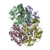
| ||||||||
|---|---|---|---|---|---|---|---|---|---|
| 1 |
| ||||||||
| Unit cell |
|
-Components
-Protein , 1 types, 4 molecules ABCD
| #1: Protein | Mass: 48655.316 Da / Num. of mol.: 4 / Fragment: RESIDUES 302-721 / Mutation: YES Source method: isolated from a genetically manipulated source Source: (gene. exp.) HOMO SAPIENS (human) / Production host:  |
|---|
-Non-polymers , 6 types, 877 molecules 


| #2: Chemical | ChemComp-ARG /  Type: L-peptide linking / Mass: 175.209 Da / Num. of mol.: 4 / Source method: obtained synthetically / Formula: C6H15N4O2 Type: L-peptide linking / Mass: 175.209 Da / Num. of mol.: 4 / Source method: obtained synthetically / Formula: C6H15N4O2#3: Chemical | ChemComp-HEM /  Mass: 616.487 Da / Num. of mol.: 4 / Source method: obtained synthetically / Formula: C34H32FeN4O4 Mass: 616.487 Da / Num. of mol.: 4 / Source method: obtained synthetically / Formula: C34H32FeN4O4#4: Chemical | ChemComp-H4B /  Mass: 241.247 Da / Num. of mol.: 4 / Source method: obtained synthetically / Formula: C9H15N5O3 Mass: 241.247 Da / Num. of mol.: 4 / Source method: obtained synthetically / Formula: C9H15N5O3#5: Chemical | ChemComp-GOL /  Mass: 92.094 Da / Num. of mol.: 7 / Source method: obtained synthetically / Formula: C3H8O3 Mass: 92.094 Da / Num. of mol.: 7 / Source method: obtained synthetically / Formula: C3H8O3#6: Chemical |  Mass: 65.409 Da / Num. of mol.: 2 / Source method: obtained synthetically / Formula: Zn Mass: 65.409 Da / Num. of mol.: 2 / Source method: obtained synthetically / Formula: Zn#7: Water | ChemComp-HOH / | Mass: 18.015 Da / Num. of mol.: 856 / Source method: isolated from a natural source / Formula: H2O |
|---|
-Details
| Nonpolymer details | L-ARGININE (LRG): SUBSTRATE OF NOS |
|---|
-Experimental details
-Experiment
| Experiment | Method: X-RAY DIFFRACTION / Number of used crystals: 1 |
|---|
- Sample preparation
| Crystal | Density Matthews: 3.22 Å3/Da / Density % sol: 61.8 % / Description: RPIM 0.446 CC ONE HALF 0.666 |
|---|---|
| Crystal grow | pH: 5 Details: 11-13% PEG3350, 50MM CITRIC ACID 50MM BIS-TRIS-PROPANE, 10% GLYCEROL, 5 MM TCEP, pH 5.0 |
-Data collection
| Diffraction | Mean temperature: 100 K |
|---|---|
| Diffraction source | Source: SYNCHROTRON / Site: SSRL  / Beamline: BL7-1 / Wavelength: 1.127 / Beamline: BL7-1 / Wavelength: 1.127 |
| Detector | Type: ADSC QUANTUM 315r / Detector: CCD / Date: Jun 1, 2013 / Details: MIRRORS |
| Radiation | Monochromator: GRAPHITE / Protocol: SINGLE WAVELENGTH / Monochromatic (M) / Laue (L): M / Scattering type: x-ray |
| Radiation wavelength | Wavelength: 1.127 Å / Relative weight: 1 |
| Reflection | Resolution: 2.03→50 Å / Num. obs: 149877 / % possible obs: 94.9 % / Observed criterion σ(I): -3 / Redundancy: 3.5 % / Biso Wilson estimate: 25.42 Å2 / Rmerge(I) obs: 0.07 / Net I/σ(I): 24 |
| Reflection shell | Resolution: 2.03→2.07 Å / Redundancy: 3.5 % / Rmerge(I) obs: 0.72 / Mean I/σ(I) obs: 1.8 / % possible all: 92 |
- Processing
| Software |
| ||||||||||||||||||||||||||||||||||||||||||||||||||||||||||||||||||||||||||||||||||||||||||||||||||||||||||||||||||||||||||||||||||||||||||||||||||||||||||||||||||||||||||||||||||||||
|---|---|---|---|---|---|---|---|---|---|---|---|---|---|---|---|---|---|---|---|---|---|---|---|---|---|---|---|---|---|---|---|---|---|---|---|---|---|---|---|---|---|---|---|---|---|---|---|---|---|---|---|---|---|---|---|---|---|---|---|---|---|---|---|---|---|---|---|---|---|---|---|---|---|---|---|---|---|---|---|---|---|---|---|---|---|---|---|---|---|---|---|---|---|---|---|---|---|---|---|---|---|---|---|---|---|---|---|---|---|---|---|---|---|---|---|---|---|---|---|---|---|---|---|---|---|---|---|---|---|---|---|---|---|---|---|---|---|---|---|---|---|---|---|---|---|---|---|---|---|---|---|---|---|---|---|---|---|---|---|---|---|---|---|---|---|---|---|---|---|---|---|---|---|---|---|---|---|---|---|---|---|---|---|
| Refinement | Method to determine structure: MOLECULAR REPLACEMENT Starting model: PDB ENTRY 1OM4 Resolution: 2.03→46.6 Å / Cor.coef. Fo:Fc: 0.965 / Cor.coef. Fo:Fc free: 0.952 / SU B: 7.285 / SU ML: 0.102 / Cross valid method: THROUGHOUT / ESU R: 0.157 / ESU R Free: 0.141 / Stereochemistry target values: MAXIMUM LIKELIHOOD Details: HYDROGENS HAVE BEEN ADDED IN THE RIDING POSITIONS. U VALUES WITH TLS ADDED. RESIDUES 344 TO 352 ARE DISORDERED IN CHAINS B AND D ARE DISORDERED
| ||||||||||||||||||||||||||||||||||||||||||||||||||||||||||||||||||||||||||||||||||||||||||||||||||||||||||||||||||||||||||||||||||||||||||||||||||||||||||||||||||||||||||||||||||||||
| Solvent computation | Ion probe radii: 0.8 Å / Shrinkage radii: 0.8 Å / VDW probe radii: 1.2 Å / Solvent model: MASK | ||||||||||||||||||||||||||||||||||||||||||||||||||||||||||||||||||||||||||||||||||||||||||||||||||||||||||||||||||||||||||||||||||||||||||||||||||||||||||||||||||||||||||||||||||||||
| Displacement parameters | Biso mean: 44.583 Å2
| ||||||||||||||||||||||||||||||||||||||||||||||||||||||||||||||||||||||||||||||||||||||||||||||||||||||||||||||||||||||||||||||||||||||||||||||||||||||||||||||||||||||||||||||||||||||
| Refinement step | Cycle: LAST / Resolution: 2.03→46.6 Å
| ||||||||||||||||||||||||||||||||||||||||||||||||||||||||||||||||||||||||||||||||||||||||||||||||||||||||||||||||||||||||||||||||||||||||||||||||||||||||||||||||||||||||||||||||||||||
| Refine LS restraints |
|