 Movie
Movie Controller

[English] 日本語
 Yorodumi
- PDB-4cyw: Structure of the A_mallard_Sweden_51_2002 H10 Avian Haemmaglutini... -
+ Open data

- Basic information
| Entry | Database: PDB / ID: 4cyw | |||||||||
|---|---|---|---|---|---|---|---|---|---|---|
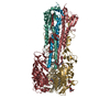
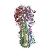
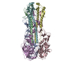
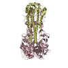
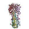
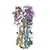
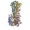
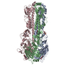
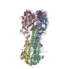
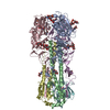
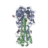
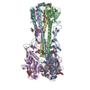
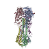
| Title | Structure of the A_mallard_Sweden_51_2002 H10 Avian Haemmaglutinin in complex with human receptor analog 6-SLN | |||||||||
 Components Components | (HEMAGGLUTININ) x 2 | |||||||||
 Keywords Keywords | VIRAL PROTEIN | |||||||||
| Function / homology |  Function and homology information Function and homology informationviral budding from plasma membrane / clathrin-dependent endocytosis of virus by host cell / host cell surface receptor binding / fusion of virus membrane with host plasma membrane / fusion of virus membrane with host endosome membrane / viral envelope / virion attachment to host cell / host cell plasma membrane / virion membrane / membrane Similarity search - Function | |||||||||
| Biological species |   INFLUENZA A VIRUS INFLUENZA A VIRUS | |||||||||
| Method |  X-RAY DIFFRACTION / SYNCHROTRON / MOLECULAR REPLACEMENT / Resolution: 2.6 Å X-RAY DIFFRACTION / SYNCHROTRON / MOLECULAR REPLACEMENT / Resolution: 2.6 Å | |||||||||
 Authors Authors | Vachieri, S.G. / Xiong, X. / Collins, P.J. / Walker, P.A. / Martin, S.R. / Haire, L.F. / McCauley, J.W. / Gamblin, S.J. / Skehel, J.J. | |||||||||
 Citation Citation | Journal: Nature / Year: 2014 Title: Receptor Binding by H10 Influenza Viruses. Authors: Vachieri, S.G. / Xiong, X. / Collins, P.J. / Walker, P.A. / Martin, S.R. / Haire, L.F. / Zhang, Y. / Mccauley, J.W. / Gamblin, S.J. / Skehel, J.J. | |||||||||
| History |
|
- Structure visualization
| Structure viewer | Molecule: MolmilJmol/JSmol |
|---|
- Downloads & links
-Download
| PDBx/mmCIF format | 4cyw.cif.gz | 603.4 KB | Display | PDBx/mmCIF format |
|---|---|---|---|---|
| PDB format | pdb4cyw.ent.gz | 504.2 KB | Display | PDB format |
| PDBx/mmJSON format | 4cyw.json.gz | Tree view | PDBx/mmJSON format | |
| Others |  Other downloads Other downloads |
-Validation report
| Arichive directory | https://data.pdbj.org/pub/pdb/validation_reports/cy/4cywftp://data.pdbj.org/pub/pdb/validation_reports/cy/4cyw | HTTPS FTP |
|---|
-Related structure data
| Related structure data | 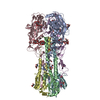 4cyvSC 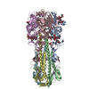 4cyzC  4cz0C 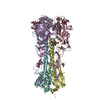 4d00C S: Starting model for refinement C: citing same article ( |
|---|---|
| Similar structure data |


-Links
 PDBj
PDBj


- Assembly
| Deposited unit | 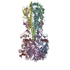
| ||||||||
|---|---|---|---|---|---|---|---|---|---|
| 1 |
| ||||||||
| Unit cell |
|
-Components
-Protein , 2 types, 6 molecules ACEBDF
| #1: Protein | Mass: 35319.750 Da / Num. of mol.: 3 / Fragment: HA1, RESIDUES 17-340 / Source method: isolated from a natural source Source: (natural) INFLUENZA A VIRUS (A/MALLARD/SWEDEN/51/2002 (H10N2))References: UniProt: E0YNJ7 #2: Protein | Mass: 19887.820 Da / Num. of mol.: 3 / Fragment: HA2, RESIDUES 341-513 / Source method: isolated from a natural source Source: (natural) INFLUENZA A VIRUS (A/MALLARD/SWEDEN/51/2002 (H10N2))References: UniProt: E0YNJ7 |
|---|
-Sugars , 5 types, 12 molecules 
| #3: Polysaccharide | 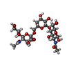  Source method: isolated from a genetically manipulated source Details: oligosaccharide / References: 6'-sialyl-N-acetyllactosamine #4: Polysaccharide | alpha-D-mannopyranose-(1-3)-[alpha-D-mannopyranose-(1-6)]beta-D-mannopyranose-(1-4)-2-acetamido-2- ...alpha-D-mannopyranose-(1-3)-[alpha-D-mannopyranose-(1-6)]beta-D-mannopyranose-(1-4)-2-acetamido-2-deoxy-beta-D-glucopyranose-(1-4)-2-acetamido-2-deoxy-beta-D-glucopyranose | Source method: isolated from a genetically manipulated source #5: Polysaccharide | beta-D-mannopyranose-(1-3)-[alpha-D-mannopyranose-(1-6)]beta-D-mannopyranose-(1-4)-2-acetamido-2- ...beta-D-mannopyranose-(1-3)-[alpha-D-mannopyranose-(1-6)]beta-D-mannopyranose-(1-4)-2-acetamido-2-deoxy-beta-D-glucopyranose-(1-4)-2-acetamido-2-deoxy-beta-D-glucopyranose | Source method: isolated from a genetically manipulated source #6: Polysaccharide | alpha-D-mannopyranose-(1-3)-beta-D-mannopyranose-(1-4)-2-acetamido-2-deoxy-beta-D-glucopyranose-(1- ...alpha-D-mannopyranose-(1-3)-beta-D-mannopyranose-(1-4)-2-acetamido-2-deoxy-beta-D-glucopyranose-(1-4)-2-acetamido-2-deoxy-beta-D-glucopyranose | Source method: isolated from a genetically manipulated source #7: Sugar | ChemComp-NAG /  Type: D-saccharide, beta linking / Mass: 221.208 Da / Num. of mol.: 6 Type: D-saccharide, beta linking / Mass: 221.208 Da / Num. of mol.: 6Source method: isolated from a genetically manipulated source Formula: C8H15NO6 |
|---|
-Non-polymers , 1 types, 285 molecules 
| #8: Water | ChemComp-HOH / Mass: 18.015 Da / Num. of mol.: 285 / Source method: isolated from a natural source / Formula: H2O |
|---|
-Details
| Has protein modification | Y |
|---|
-Experimental details
-Experiment
| Experiment | Method: X-RAY DIFFRACTION / Number of used crystals: 1 |
|---|
- Sample preparation
| Crystal | Density Matthews: 3.45 Å3/Da / Density % sol: 64.41 % / Description: NONE |
|---|---|
| Crystal grow | Details: 18% PEG 2000MME AND 0.1 M BICINE PH 9.0 |
-Data collection
| Diffraction | Mean temperature: 100 K |
|---|---|
| Diffraction source | Source: SYNCHROTRON / Site: Diamond  / Beamline: I04 / Wavelength: 0.92 / Beamline: I04 / Wavelength: 0.92 |
| Detector | Type: DECTRIS PILATUS / Detector: PIXEL |
| Radiation | Protocol: SINGLE WAVELENGTH / Monochromatic (M) / Laue (L): M / Scattering type: x-ray |
| Radiation wavelength | Wavelength: 0.92 Å / Relative weight: 1 |
| Reflection | Resolution: 2.6→48.85 Å / Num. obs: 67905 / % possible obs: 99.1 % / Observed criterion σ(I): 1.8 / Redundancy: 3.6 % / Rmerge(I) obs: 0.07 / Net I/σ(I): 11.8 |
| Reflection shell | Resolution: 2.6→2.66 Å / Redundancy: 3.7 % / Rmerge(I) obs: 0.69 / Mean I/σ(I) obs: 1.8 / % possible all: 99.8 |
- Processing
| Software |
| ||||||||||||||||||||||||||||||||||||||||||||||||||||||||||||||||||||||||||||||||||||||||||||||||||||||||||||||||||||||||||||||||||||||||||||||||||||||||||||||||||||||||||||||||||||||
|---|---|---|---|---|---|---|---|---|---|---|---|---|---|---|---|---|---|---|---|---|---|---|---|---|---|---|---|---|---|---|---|---|---|---|---|---|---|---|---|---|---|---|---|---|---|---|---|---|---|---|---|---|---|---|---|---|---|---|---|---|---|---|---|---|---|---|---|---|---|---|---|---|---|---|---|---|---|---|---|---|---|---|---|---|---|---|---|---|---|---|---|---|---|---|---|---|---|---|---|---|---|---|---|---|---|---|---|---|---|---|---|---|---|---|---|---|---|---|---|---|---|---|---|---|---|---|---|---|---|---|---|---|---|---|---|---|---|---|---|---|---|---|---|---|---|---|---|---|---|---|---|---|---|---|---|---|---|---|---|---|---|---|---|---|---|---|---|---|---|---|---|---|---|---|---|---|---|---|---|---|---|---|---|
| Refinement | Method to determine structure: MOLECULAR REPLACEMENT Starting model: PDB ENTRY 4CYV Resolution: 2.6→107.72 Å / Cor.coef. Fo:Fc: 0.948 / Cor.coef. Fo:Fc free: 0.918 / SU B: 30.236 / SU ML: 0.294 / Cross valid method: THROUGHOUT / ESU R: 0.481 / ESU R Free: 0.292 / Stereochemistry target values: MAXIMUM LIKELIHOOD Details: HYDROGENS HAVE BEEN ADDED IN THE RIDING POSITIONS. U VALUES WITH TLS ADDED
| ||||||||||||||||||||||||||||||||||||||||||||||||||||||||||||||||||||||||||||||||||||||||||||||||||||||||||||||||||||||||||||||||||||||||||||||||||||||||||||||||||||||||||||||||||||||
| Solvent computation | Ion probe radii: 0.8 Å / Shrinkage radii: 0.8 Å / VDW probe radii: 1.2 Å / Solvent model: MASK | ||||||||||||||||||||||||||||||||||||||||||||||||||||||||||||||||||||||||||||||||||||||||||||||||||||||||||||||||||||||||||||||||||||||||||||||||||||||||||||||||||||||||||||||||||||||
| Displacement parameters | Biso mean: 60.888 Å2
| ||||||||||||||||||||||||||||||||||||||||||||||||||||||||||||||||||||||||||||||||||||||||||||||||||||||||||||||||||||||||||||||||||||||||||||||||||||||||||||||||||||||||||||||||||||||
| Refinement step | Cycle: LAST / Resolution: 2.6→107.72 Å
| ||||||||||||||||||||||||||||||||||||||||||||||||||||||||||||||||||||||||||||||||||||||||||||||||||||||||||||||||||||||||||||||||||||||||||||||||||||||||||||||||||||||||||||||||||||||
| Refine LS restraints |
|