 Movie
Movie Controller

[English] 日本語
 Yorodumi
- PDB-4cpk: Crystal structure of PBP2a double clinical mutant N146K-E150K fro... -
+ Open data

- Basic information
| Entry | Database: PDB / ID: 4cpk | ||||||
|---|---|---|---|---|---|---|---|
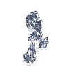
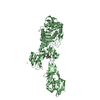
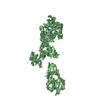
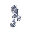
| Title | Crystal structure of PBP2a double clinical mutant N146K-E150K from MRSA | ||||||
 Components Components | Penicillin binding protein 2 prime | ||||||
 Keywords Keywords | HYDROLASE / PENICILLIN-BINDING PROTEIN / MRSA / ALLOSTERIC SITE | ||||||
| Function / homology |  Function and homology information Function and homology informationpeptidoglycan L,D-transpeptidase activity / penicillin binding / cell wall organization / response to antibiotic / plasma membrane Similarity search - Function | ||||||
| Biological species |   Staphylococcus aureus (bacteria) Staphylococcus aureus (bacteria) | ||||||
| Method |  X-RAY DIFFRACTION / SYNCHROTRON / MOLECULAR REPLACEMENT / Resolution: 2.35 Å X-RAY DIFFRACTION / SYNCHROTRON / MOLECULAR REPLACEMENT / Resolution: 2.35 Å | ||||||
 Authors Authors | Otero, L.H. / Rojas-Altuve, A. / Hermoso, J.A. | ||||||
 Citation Citation | Journal: J.Am.Chem.Soc. / Year: 2014 Title: Disruption of Allosteric Response as an Unprecedented Mechanism of Resistance to Antibiotics. Authors: Fishovitz, J. / Rojas-Altuve, A. / Otero, L.H. / Dawley, M. / Carrasco-Lopez, C. / Chang, M. / Hermoso, J.A. / Mobashery, S. | ||||||
| History |
|
- Structure visualization
| Structure viewer | Molecule: MolmilJmol/JSmol |
|---|
- Downloads & links
-Download
| PDBx/mmCIF format | 4cpk.cif.gz | 538.1 KB | Display | PDBx/mmCIF format |
|---|---|---|---|---|
| PDB format | pdb4cpk.ent.gz | 441.5 KB | Display | PDB format |
| PDBx/mmJSON format | 4cpk.json.gz | Tree view | PDBx/mmJSON format | |
| Others |  Other downloads Other downloads |
-Validation report
| Arichive directory | https://data.pdbj.org/pub/pdb/validation_reports/cp/4cpkftp://data.pdbj.org/pub/pdb/validation_reports/cp/4cpk | HTTPS FTP |
|---|
-Related structure data
| Related structure data |  4bl2C  4bl3C  3zg0S S: Starting model for refinement C: citing same article ( |
|---|---|
| Similar structure data |


-Links
 PDBj
PDBj

- Assembly
| Deposited unit | 
| ||||||||
|---|---|---|---|---|---|---|---|---|---|
| 1 | 
| ||||||||
| 2 | 
| ||||||||
| Unit cell |
|
-Components
| #1: Protein | Mass: 73430.984 Da / Num. of mol.: 2 / Fragment: RESIDUES 26-668 / Mutation: YES,N146K,E150K Source method: isolated from a genetically manipulated source Source: (gene. exp.) Staphylococcus aureus (strain Mu50 / ATCC 700699) (bacteria)Strain: Mu50 / ATCC 700699 / Gene: mecA, SAV0041 / Plasmid: pET24D / Production host: #2: Chemical | ChemComp-CD /   Mass: 112.411 Da / Num. of mol.: 11 / Source method: obtained synthetically / Formula: Cd Mass: 112.411 Da / Num. of mol.: 11 / Source method: obtained synthetically / Formula: Cd#3: Chemical | ChemComp-CL /   Mass: 35.453 Da / Num. of mol.: 4 / Source method: obtained synthetically / Formula: Cl Mass: 35.453 Da / Num. of mol.: 4 / Source method: obtained synthetically / Formula: Cl#4: Sugar |   Type: D-saccharide, beta linking / Mass: 251.234 Da / Num. of mol.: 2 / Source method: obtained synthetically / Formula: C9H17NO7 Type: D-saccharide, beta linking / Mass: 251.234 Da / Num. of mol.: 2 / Source method: obtained synthetically / Formula: C9H17NO7#5: Water | ChemComp-HOH / |  Mass: 18.015 Da / Num. of mol.: 764 / Source method: isolated from a natural source / Formula: H2O Mass: 18.015 Da / Num. of mol.: 764 / Source method: isolated from a natural source / Formula: H2O |
|---|
-Experimental details
-Experiment
| Experiment | Method: X-RAY DIFFRACTION |
|---|
- Sample preparation
| Crystal | Density Matthews: 2.63 Å3/Da / Density % sol: 53.28 % / Description: NONE |
|---|---|
| Crystal grow | pH: 7 / Details: pH 7 |
-Data collection
| Diffraction | Mean temperature: 100 K |
|---|---|
| Diffraction source | Source: SYNCHROTRON / Site: ESRF  / Beamline: ID23-2 / Wavelength: 0.8729 / Beamline: ID23-2 / Wavelength: 0.8729 |
| Detector | Type: MARRESEARCH / Detector: CCD |
| Radiation | Protocol: SINGLE WAVELENGTH / Monochromatic (M) / Laue (L): M / Scattering type: x-ray |
| Radiation wavelength | Wavelength: 0.8729 Å / Relative weight: 1 |
| Reflection | Resolution: 2.35→46.64 Å / Num. obs: 65572 / % possible obs: 100 % / Observed criterion σ(I): 2 / Redundancy: 8.3 % / Biso Wilson estimate: 40.68 Å2 / Rmerge(I) obs: 0.16 / Net I/σ(I): 11.6 |
| Reflection shell | Resolution: 2.35→2.48 Å / Redundancy: 8.4 % / Rmerge(I) obs: 0.94 / Mean I/σ(I) obs: 2.3 / % possible all: 100 |
- Processing
| Software |
| ||||||||||||||||||||||||||||||||||||||||||||||||||||||||||||||||||||||||||||||||||||||||||||||||||||||||||||||||||
|---|---|---|---|---|---|---|---|---|---|---|---|---|---|---|---|---|---|---|---|---|---|---|---|---|---|---|---|---|---|---|---|---|---|---|---|---|---|---|---|---|---|---|---|---|---|---|---|---|---|---|---|---|---|---|---|---|---|---|---|---|---|---|---|---|---|---|---|---|---|---|---|---|---|---|---|---|---|---|---|---|---|---|---|---|---|---|---|---|---|---|---|---|---|---|---|---|---|---|---|---|---|---|---|---|---|---|---|---|---|---|---|---|---|---|---|
| Refinement | Method to determine structure: MOLECULAR REPLACEMENT Starting model: PDB ENTRY 3ZG0 Resolution: 2.35→46.64 Å / Cor.coef. Fo:Fc: 0.9387 / Cor.coef. Fo:Fc free: 0.9077 / SU R Cruickshank DPI: 0.306 / Cross valid method: THROUGHOUT / σ(F): 0 / SU R Blow DPI: 0.33 / SU Rfree Blow DPI: 0.225 / SU Rfree Cruickshank DPI: 0.222
| ||||||||||||||||||||||||||||||||||||||||||||||||||||||||||||||||||||||||||||||||||||||||||||||||||||||||||||||||||
| Displacement parameters | Biso mean: 47.43 Å2
| ||||||||||||||||||||||||||||||||||||||||||||||||||||||||||||||||||||||||||||||||||||||||||||||||||||||||||||||||||
| Refine analyze | Luzzati coordinate error obs: 0.306 Å | ||||||||||||||||||||||||||||||||||||||||||||||||||||||||||||||||||||||||||||||||||||||||||||||||||||||||||||||||||
| Refinement step | Cycle: LAST / Resolution: 2.35→46.64 Å
| ||||||||||||||||||||||||||||||||||||||||||||||||||||||||||||||||||||||||||||||||||||||||||||||||||||||||||||||||||
| Refine LS restraints |
| ||||||||||||||||||||||||||||||||||||||||||||||||||||||||||||||||||||||||||||||||||||||||||||||||||||||||||||||||||
| LS refinement shell | Resolution: 2.35→2.41 Å / Total num. of bins used: 20
| ||||||||||||||||||||||||||||||||||||||||||||||||||||||||||||||||||||||||||||||||||||||||||||||||||||||||||||||||||
| Refinement TLS params. | Method: refined / Refine-ID: X-RAY DIFFRACTION
|
