Mass: 18.015 Da / Num. of mol.: 83 / Source method: isolated from a natural source / Formula: H2O
Has protein modification
Y
-
Experimental details
-
Experiment
Experiment
Method: X-RAY DIFFRACTION / Number of used crystals: 1
-
Sample preparation
Crystal
Density Matthews: 2.25 Å3/Da / Density % sol: 45.36 % / Description: NONE
Crystal grow
Method: vapor diffusion, sitting drop Details: 0.1 M MAGNESIUM CHLORIDE, 0.1M SODIUM CHLORIDE, 0.1 M MES PH6.5, 12% PEG 4000, 0.05 M LACTOSE, VAPOR DIFFUSION IN SITTING DROP, SOAKED WITH GALB1-4GLCNAC
Resolution: 2.6→41 Å / Cor.coef. Fo:Fc: 0.948 / Cor.coef. Fo:Fc free: 0.912 / SU B: 16.66 / SU ML: 0.199 / Cross valid method: THROUGHOUT / ESU R: 0.55 / ESU R Free: 0.294 / Stereochemistry target values: MAXIMUM LIKELIHOOD Details: HYDROGENS HAVE BEEN ADDED IN THE RIDING POSITIONS. U VALUES WITH TLS ADDED. PART OF LOOP L1 NOT DEFINED IN ELECTRON DENSITY, LYS A 76 - GLY A 107
Rfactor
Num. reflection
% reflection
Selection details
Rfree
0.24228
492
5.1 %
RANDOM
Rwork
0.18086
-
-
-
obs
0.18392
9180
99.86 %
-
Solvent computation
Ion probe radii: 0.8 Å / Shrinkage radii: 0.8 Å / VDW probe radii: 1.2 Å / Solvent model: MASK
 Movie
Movie Controller

 Yorodumi
 Open data
 Basic information
 Components
 Keywords
 Function and homology information
 CANDIDA GLABRATA (fungus)
 X-RAY DIFFRACTION /
  Authors
 Citation
 Structure visualization
 Downloads & links
 Other downloads


 PDBj
PDBj
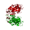
 Assembly


 Mass: 40.078 Da / Num. of mol.: 1 / Source method: obtained synthetically / Formula: Ca
 Mass: 35.453 Da / Num. of mol.: 1 / Source method: obtained synthetically / Formula: Cl
 Mass: 18.015 Da / Num. of mol.: 83 / Source method: isolated from a natural source / Formula: H2O
 Sample preparation
 / Beamline: 14.1 / Wavelength: 0.918409
  Processing