Monochromator: SI FILTER / Protocol: SINGLE WAVELENGTH / Monochromatic (M) / Laue (L): M / Scattering type: x-ray
Radiation wavelength
Wavelength: 1.5418 Å / Relative weight: 1
Reflection
Resolution: 1.95→42.48 Å / Num. obs: 202165 / % possible obs: 99.4 % / Observed criterion σ(I): 0 / Redundancy: 3.5 % / Rmerge(I) obs: 0.06 / Net I/σ(I): 11.1
Reflection shell
Resolution: 1.95→2 Å / Redundancy: 2.6 % / Rmerge(I) obs: 0.12 / Mean I/σ(I) obs: 4.3 / % possible all: 94.9
-
Processing
Software
Name
Version
Classification
REFMAC
5.8.0049
refinement
iMOSFLM
datareduction
Aimless
datascaling
MOLREP
phasing
Refinement
Method to determine structure: MOLECULAR REPLACEMENT Starting model: KYNURENINE FORMAMIDASE FROM BURKHOLDERIA CENOCEPACIA. TO BE SUBMITED Resolution: 1.95→83.76 Å / Cor.coef. Fo:Fc: 0.95 / Cor.coef. Fo:Fc free: 0.927 / SU B: 3.595 / SU ML: 0.102 / Cross valid method: THROUGHOUT / ESU R: 0.188 / ESU R Free: 0.152 / Stereochemistry target values: MAXIMUM LIKELIHOOD / Details: HYDROGENS HAVE BEEN ADDED IN THE RIDING POSITIONS.
Rfactor
Num. reflection
% reflection
Selection details
Rfree
0.20663
2954
5.1 %
RANDOM
Rwork
0.17145
-
-
-
obs
0.17323
55358
99.37 %
-
Solvent computation
Ion probe radii: 0.8 Å / Shrinkage radii: 0.8 Å / VDW probe radii: 1.2 Å / Solvent model: MASK
 Movie
Movie Controller

 Yorodumi
 Open data
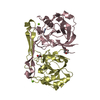
 Basic information
 Components
 Keywords
 Function and homology information
 X-RAY DIFFRACTION /
  Authors
 Citation
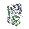
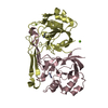
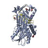
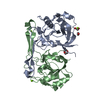
 Structure visualization
 Downloads & links
 Other downloads
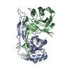


 PDBj
PDBj Assembly


 Mass: 65.409 Da / Num. of mol.: 8 / Source method: obtained synthetically / Formula: Zn
 Mass: 24.305 Da / Num. of mol.: 5 / Source method: obtained synthetically / Formula: Mg
 Mass: 88.105 Da / Num. of mol.: 2 / Source method: obtained synthetically / Formula: C4H8O2
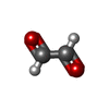
 Mass: 58.036 Da / Num. of mol.: 3 / Source method: obtained synthetically / Formula: C2H2O2
 Sample preparation
 Processing