[English] 日本語
Yorodumi
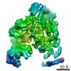
- PDB-4ci6: Mechanisms of crippling actin-dependent phagocytosis by YopO -

+
Open data


ID or keywords:

Loading...

-
Basic information

Entry
Database: PDB / ID: 4ci6
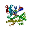
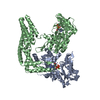
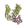
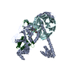
TitleMechanisms of crippling actin-dependent phagocytosis by YopO
Components
  • ACTIN
  • PROTEIN KINASE YOPO
KeywordsTRANSFERASE/STRUCTURAL PROTEIN / TRANSFERASE-STRUCTURAL PROTEIN COMPLEX / BUBONIC PLAGUE
Function / homology
Function and homology information


non-specific serine/threonine protein kinase / phosphorylation / protein serine/threonine kinase activity / ATP binding
Similarity search - Function
Serine/threonine protein kinase, yersinia-type / YpkA, Rac1-binding domain / Rac1-binding domain, C-terminal / Rac1-binding domain, N-terminal / Rac1-binding domain / ATPase, substrate binding domain, subdomain 4 / Actin; Chain A, domain 4 / ATPase, nucleotide binding domain / Actins signature 1. / Actin, conserved site ...Serine/threonine protein kinase, yersinia-type / YpkA, Rac1-binding domain / Rac1-binding domain, C-terminal / Rac1-binding domain, N-terminal / Rac1-binding domain / ATPase, substrate binding domain, subdomain 4 / Actin; Chain A, domain 4 / ATPase, nucleotide binding domain / Actins signature 1. / Actin, conserved site / Actins signature 2. / Actin/actin-like conserved site / Actins and actin-related proteins signature. / Actin / Actin family / Actin / ATPase, nucleotide binding domain / Nucleotidyltransferase; domain 5 / Serine/threonine-protein kinase, active site / Serine/Threonine protein kinases active-site signature. / Protein kinase domain / Serine/Threonine protein kinases, catalytic domain / Protein kinase domain profile. / Protein kinase domain / Protein kinase-like domain superfamily / Alpha-Beta Complex / 2-Layer Sandwich / Alpha Beta
Similarity search - Domain/homology
ADENOSINE-5'-TRIPHOSPHATE / Actin / YopO
Similarity search - Component
Biological speciesYERSINIA ENTEROCOLITICA (bacteria)
SPODOPTERA FRUGIPERDA (fall armyworm)
MethodX-RAY DIFFRACTION / SYNCHROTRON / MOLECULAR REPLACEMENT / Resolution: 2.651 Å
AuthorsLee, W.L. / Grimes, J.M. / Robinson, R.C.
CitationJournal: Nat.Struct.Mol.Biol. / Year: 2015
Title: Yersinia Effector Yopo Uses Actin as Bait to Phosphorylate Proteins that Regulate Actin Polymerization.
Authors: Lee, W.L. / Grimes, J.M. / Robinson, R.C.
History
DepositionDec 6, 2013Deposition site: PDBE / Processing site: PDBE
Revision 1.0Jan 28, 2015Provider: repository / Type: Initial release
Revision 1.1Feb 11, 2015Group: Database references
Revision 1.2Mar 4, 2015Group: Database references
Revision 1.3Mar 18, 2015Group: Database references
Revision 1.4Dec 20, 2023Group: Data collection / Database references ...Data collection / Database references / Derived calculations / Other / Refinement description
Category: chem_comp_atom / chem_comp_bond ...chem_comp_atom / chem_comp_bond / database_2 / pdbx_database_status / pdbx_initial_refinement_model / pdbx_struct_conn_angle / struct_conn / struct_site
Item: _database_2.pdbx_DOI / _database_2.pdbx_database_accession ..._database_2.pdbx_DOI / _database_2.pdbx_database_accession / _pdbx_database_status.status_code_sf / _pdbx_struct_conn_angle.ptnr1_auth_seq_id / _pdbx_struct_conn_angle.ptnr3_auth_seq_id / _pdbx_struct_conn_angle.value / _struct_conn.pdbx_dist_value / _struct_conn.ptnr2_auth_seq_id / _struct_site.pdbx_auth_asym_id / _struct_site.pdbx_auth_comp_id / _struct_site.pdbx_auth_seq_id

-
Structure visualization

Structure viewerMolecule:
MolmilJmol/JSmol

Downloads & links

-
Assembly

Deposited unit
A: ACTIN
B: PROTEIN KINASE YOPO
hetero molecules


Theoretical massNumber of molelcules
Total (without water)114,0804
Polymers113,5322
Non-polymers5472
Water3,081171
1


  • Idetical with deposited unit
  • defined by author&software
TypeNameSymmetry operationNumber
identity operation1_555x,y,z1
Buried area5150 Å2
ΔGint-25.9 kcal/mol
Surface area41130 Å2
MethodPISA
Unit cell
Length a, b, c (Å)70.799, 121.850, 89.339
Angle α, β, γ (deg.)90.00, 94.87, 90.00
Int Tables number4
Space group name H-MP1211

-
Components

#1: Protein ACTIN /


Mass: 41755.617 Da / Num. of mol.: 1 / Fragment: RESIDUES 89-729 / Source method: isolated from a natural source / Source: (natural) SPODOPTERA FRUGIPERDA (fall armyworm) / References: UniProt: G3CKA6
#2: Protein PROTEIN KINASE YOPO


Mass: 71776.875 Da / Num. of mol.: 1 / Mutation: YES
Source method: isolated from a genetically manipulated source
Source: (gene. exp.) YERSINIA ENTEROCOLITICA (bacteria) / Production host: ESCHERICHIA COLI (E. coli) / References: UniProt: Q93KQ6
#3: Chemical ChemComp-ATP / ADENOSINE-5'-TRIPHOSPHATE / Adenosine triphosphate


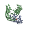
Mass: 507.181 Da / Num. of mol.: 1 / Source method: obtained synthetically / Formula: C10H16N5O13P3 / Comment: ATP, energy-carrying molecule*YM
#4: Chemical ChemComp-CA / CALCIUM ION


Mass: 40.078 Da / Num. of mol.: 1 / Source method: obtained synthetically / Formula: Ca
#5: Water ChemComp-HOH / water / Water


Mass: 18.015 Da / Num. of mol.: 171 / Source method: isolated from a natural source / Formula: H2O

-
Experimental details

-
Experiment

ExperimentMethod: X-RAY DIFFRACTION / Number of used crystals: 1

-
Sample preparation

CrystalDensity Matthews: 3.4 Å3/Da / Density % sol: 64 % / Description: NONE
Crystal growpH: 7.5 / Details: 30% PEG400, 0.1M HEPES PH7.5, 0.2M SODIUM CHLORIDE

-
Data collection

DiffractionMean temperature: 110 K
Diffraction sourceSource: SYNCHROTRON / Site: NSRRC / Beamline: BL13B1 / Wavelength: 1
DetectorType: ADSC QUANTUM 315 / Detector: CCD / Details: TOROIDAL FOCUSING MIRROR
RadiationMonochromator: LN2-COOLED, FIXED-EXIT DOUBLE CRYSTAL MONOCHROMATOR
Protocol: SINGLE WAVELENGTH / Monochromatic (M) / Laue (L): M / Scattering type: x-ray
Radiation wavelengthWavelength: 1 Å / Relative weight: 1
ReflectionResolution: 2.65→20 Å / Num. obs: 41770 / % possible obs: 95.9 % / Observed criterion σ(I): 0 / Redundancy: 3.6 % / Rmerge(I) obs: 0.1 / Net I/σ(I): 12.75
Reflection shellResolution: 2.65→2.7 Å / Redundancy: 3.2 % / Rmerge(I) obs: 0.47 / Mean I/σ(I) obs: 2.65 / % possible all: 75.1

-
Processing

Software
NameVersionClassification
PHENIX(PHENIX.REFINE: 1.8.4_1496)refinement
HKL-2000data reduction
SCALEPACKdata scaling
PHASERphasing
RefinementMethod to determine structure: MOLECULAR REPLACEMENT
Starting model: PDB ENTRIES 3HBT, 2H7O
Resolution: 2.651→20.022 Å / SU ML: 0.28 / σ(F): 1.34 / Phase error: 25.78 / Stereochemistry target values: ML
RfactorNum. reflection% reflection
Rfree0.2201 2107 5 %
Rwork0.1856 --
obs0.1873 41765 95.45 %
Solvent computationShrinkage radii: 1 Å / VDW probe radii: 1.2 Å / Solvent model: FLAT BULK SOLVENT MODEL
Refinement stepCycle: LAST / Resolution: 2.651→20.022 Å
ProteinNucleic acidLigandSolventTotal
Num. atoms7315 0 32 171 7518
Refine LS restraints
Refine-IDTypeDev idealNumber
X-RAY DIFFRACTIONf_bond_d0.0027490
X-RAY DIFFRACTIONf_angle_d0.60510142
X-RAY DIFFRACTIONf_dihedral_angle_d12.9172793
X-RAY DIFFRACTIONf_chiral_restr0.0231145
X-RAY DIFFRACTIONf_plane_restr0.0031301
LS refinement shell
Resolution (Å)Rfactor RfreeNum. reflection RfreeRfactor RworkNum. reflection RworkRefine-ID% reflection obs (%)
2.6508-2.71230.28621180.25441931X-RAY DIFFRACTION71
2.7123-2.780.30231170.24952262X-RAY DIFFRACTION82
2.78-2.85490.32241310.25452420X-RAY DIFFRACTION88
2.8549-2.93870.32441240.25722619X-RAY DIFFRACTION94
2.9387-3.03320.29661320.2352731X-RAY DIFFRACTION99
3.0332-3.14120.2911490.23262769X-RAY DIFFRACTION100
3.1412-3.26650.24251370.2192752X-RAY DIFFRACTION100
3.2665-3.41450.24721480.2152765X-RAY DIFFRACTION100
3.4145-3.59350.22161530.19132761X-RAY DIFFRACTION100
3.5935-3.81720.20491380.18122763X-RAY DIFFRACTION100
3.8172-4.10960.20841420.15722773X-RAY DIFFRACTION100
4.1096-4.51880.17131440.14332765X-RAY DIFFRACTION100
4.5188-5.16290.17851620.15172767X-RAY DIFFRACTION100
5.1629-6.46810.21211540.18292781X-RAY DIFFRACTION100
6.4681-20.0230.16841580.14662799X-RAY DIFFRACTION100
Refinement TLS params.Method: refined / Origin x: 41.406 Å / Origin y: 26.8596 Å / Origin z: 12.1554 Å
111213212223313233
T0.1993 Å2-0.0061 Å2-0.0147 Å2-0.1932 Å2-0.0241 Å2--0.2136 Å2
L0.2312 °20 °20.0464 °2-0.8529 °2-0.1863 °2--0.4696 °2
S0.0249 Å °-0.0258 Å °0 Å °-0.0217 Å °-0.0027 Å °0.0321 Å °0.0639 Å °0.0111 Å °-0.0153 Å °
Refinement TLS groupSelection details: ALL

+
About Yorodumi

-
News

-
Feb 9, 2022. New format data for meta-information of EMDB entries

New format data for meta-information of EMDB entries

  • Version 3 of the EMDB header file is now the official format.
  • The previous official version 1.9 will be removed from the archive.

Related info.:EMDB header

External links:wwPDB to switch to version 3 of the EMDB data model

-
Aug 12, 2020. Covid-19 info

Covid-19 info

URL: https://pdbj.org/emnavi/covid19.php

New page: Covid-19 featured information page in EM Navigator.

Related info.:Covid-19 info / Mar 5, 2020. Novel coronavirus structure data

+
Mar 5, 2020. Novel coronavirus structure data

Novel coronavirus structure data

Related info.:Yorodumi Speices / Aug 12, 2020. Covid-19 info

External links:COVID-19 featured content - PDBj / Molecule of the Month (242):Coronavirus Proteases

+
Jan 31, 2019. EMDB accession codes are about to change! (news from PDBe EMDB page)

EMDB accession codes are about to change! (news from PDBe EMDB page)

  • The allocation of 4 digits for EMDB accession codes will soon come to an end. Whilst these codes will remain in use, new EMDB accession codes will include an additional digit and will expand incrementally as the available range of codes is exhausted. The current 4-digit format prefixed with “EMD-” (i.e. EMD-XXXX) will advance to a 5-digit format (i.e. EMD-XXXXX), and so on. It is currently estimated that the 4-digit codes will be depleted around Spring 2019, at which point the 5-digit format will come into force.
  • The EM Navigator/Yorodumi systems omit the EMD- prefix.

Related info.:Q: What is EMD? / ID/Accession-code notation in Yorodumi/EM Navigator

External links:EMDB Accession Codes are Changing Soon! / Contact to PDBj

+
Jul 12, 2017. Major update of PDB

Major update of PDB

  • wwPDB released updated PDB data conforming to the new PDBx/mmCIF dictionary.
  • This is a major update changing the version number from 4 to 5, and with Remediation, in which all the entries are updated.
  • In this update, many items about electron microscopy experimental information are reorganized (e.g. em_software).
  • Now, EM Navigator and Yorodumi are based on the updated data.

External links:wwPDB Remediation / Enriched Model Files Conforming to OneDep Data Standards Now Available in the PDB FTP Archive

-
Yorodumi

Thousand views of thousand structures

  • Yorodumi is a browser for structure data from EMDB, PDB, SASBDB, etc.
  • This page is also the successor to EM Navigator detail page, and also detail information page/front-end page for Omokage search.
  • The word "yorodu" (or yorozu) is an old Japanese word meaning "ten thousand". "mi" (miru) is to see.

Related info.:EMDB / PDB / SASBDB / Comparison of 3 databanks / Yorodumi Search / Aug 31, 2016. New EM Navigator & Yorodumi / Yorodumi Papers / Jmol/JSmol / Function and homology information / Changes in new EM Navigator and Yorodumi

Read more