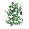
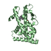
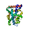
Entry Database : PDB / ID : 4ci4Title Structural basis for GL479 a dual Peroxisome Proliferator-Activated Receptor alpha agonist PEROXISOME PROLIFERATOR-ACTIVATED RECEPTOR ALPHA Keywords / / / / Function / homology Function Domain/homology Component
/ / / / / / / / / / / / / / / / / / / / / / / / / / / / / / / / / / / / / / / / / / / / / / / / / / / / / / / / / / / / / / / / / / / / / / / / / / / / / / / / / / / / / / / / / / / / / / / / / / / / / / / / / / / / / / / / / / / / / / / / / / / / Biological species HOMO SAPIENS (human)Method / / / Resolution : 2.302 Å Authors Santos, J.C. / Bernardes, A. / Polikarpov, I. Journal : J. Struct. Biol. / Year : 2015Title : Different binding and recognition modes of GL479, a dual agonist of Peroxisome Proliferator-Activated Receptor alpha / gamma.Authors : dos Santos, J.C. / Bernardes, A. / Giampietro, L. / Ammazzalorso, A. / De Filippis, B. / Amoroso, R. / Polikarpov, I. History Deposition Dec 5, 2013 Deposition site / Processing site Revision 1.0 Dec 24, 2014 Provider / Type Revision 1.1 Aug 5, 2015 Group Revision 1.2 Sep 9, 2015 Group Revision 1.3 Jan 16, 2019 Group / Database references / Category / citation_authorItem _citation.journal_abbrev / _citation.journal_id_ISSN ... _citation.journal_abbrev / _citation.journal_id_ISSN / _citation.page_last / _citation.pdbx_database_id_DOI / _citation.title / _citation_author.name Revision 1.4 Dec 20, 2023 Group Data collection / Database references ... Data collection / Database references / Derived calculations / Other / Refinement description Category chem_comp_atom / chem_comp_bond ... chem_comp_atom / chem_comp_bond / database_2 / pdbx_database_status / pdbx_initial_refinement_model / struct_site Item _database_2.pdbx_DOI / _database_2.pdbx_database_accession ... _database_2.pdbx_DOI / _database_2.pdbx_database_accession / _pdbx_database_status.status_code_sf / _struct_site.pdbx_auth_asym_id / _struct_site.pdbx_auth_comp_id / _struct_site.pdbx_auth_seq_id
Show all Show less
 Movie
Movie Controller

 Yorodumi
 Open data
 Basic information
 Components
 Keywords
 Function and homology information
 HOMO SAPIENS (human)
 X-RAY DIFFRACTION /
  Authors
 Citation
 Structure visualization
 Downloads & links
 Other downloads


 PDBj
PDBj


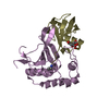
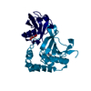
 Assembly

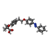
 Mass: 404.458 Da / Num. of mol.: 1 / Source method: obtained synthetically / Formula: C24H24N2O4
 Mass: 18.015 Da / Num. of mol.: 52 / Source method: isolated from a natural source / Formula: H2O
 Sample preparation
 / Beamline: W01B-MX2 / Wavelength: 1.459
  Processing