 Movie
Movie Controller

[English] 日本語
 Yorodumi
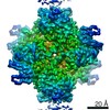
- PDB-4chv: The electron crystallography structure of the cAMP-bound potassiu... -
+ Open data

- Basic information
| Entry | Database: PDB / ID: 4chv | ||||||
|---|---|---|---|---|---|---|---|
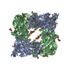
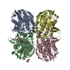
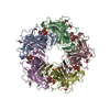
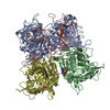
| Title | The electron crystallography structure of the cAMP-bound potassium channel MloK1 | ||||||
 Components Components | CYCLIC NUCLEOTIDE-GATED POTASSIUM CHANNEL MLL3241 | ||||||
 Keywords Keywords | TRANSPORT / 2DX / VOLTAGE GATED POTASSIUM CHANNEL / CNBD / 2D CRYSTAL | ||||||
| Function / homology |  Function and homology information Function and homology informationintracellularly cyclic nucleotide-activated monoatomic cation channel activity / potassium channel activity / cAMP binding / protein-containing complex binding / identical protein binding / plasma membrane Similarity search - Function | ||||||
| Biological species |  MESORHIZOBIUM LOTI (bacteria) MESORHIZOBIUM LOTI (bacteria) | ||||||
| Method | ELECTRON CRYSTALLOGRAPHY / electron crystallography / cryo EM / Resolution: 7 Å | ||||||
 Authors Authors | Kowal, J. / Chami, M. / Baumgartner, P. / Arheit, M. / Chiu, P.L. / Rangl, M. / Scheuring, S. / Schroeder, G.F. / Nimigean, C.M. / Stahlberg, H. | ||||||
 Citation Citation | Journal: Nat Commun / Year: 2014 Title: Ligand-induced structural changes in the cyclic nucleotide-modulated potassium channel MloK1. Authors: Julia Kowal / Mohamed Chami / Paul Baumgartner / Marcel Arheit / Po-Lin Chiu / Martina Rangl / Simon Scheuring / Gunnar F Schröder / Crina M Nimigean / Henning Stahlberg /     Abstract: Cyclic nucleotide-modulated ion channels are important for signal transduction and pacemaking in eukaryotes. The molecular determinants of ligand gating in these channels are still unknown, mainly ...Cyclic nucleotide-modulated ion channels are important for signal transduction and pacemaking in eukaryotes. The molecular determinants of ligand gating in these channels are still unknown, mainly because of a lack of direct structural information. Here we report ligand-induced conformational changes in full-length MloK1, a cyclic nucleotide-modulated potassium channel from the bacterium Mesorhizobium loti, analysed by electron crystallography and atomic force microscopy. Upon cAMP binding, the cyclic nucleotide-binding domains move vertically towards the membrane, and directly contact the S1-S4 voltage sensor domains. This is accompanied by a significant shift and tilt of the voltage sensor domain helices. In both states, the inner pore-lining helices are in an 'open' conformation. We propose a mechanism in which ligand binding can favour pore opening via a direct interaction between the cyclic nucleotide-binding domains and voltage sensors. This offers a simple mechanistic hypothesis for the coupling between ligand gating and voltage sensing in eukaryotic HCN channels. | ||||||
| History |
|
- Structure visualization
| Movie |
Movie viewer |
|---|---|
| Structure viewer | Molecule: MolmilJmol/JSmol |
- Downloads & links
-Download
| PDBx/mmCIF format | 4chv.cif.gz | 254.4 KB | Display | PDBx/mmCIF format |
|---|---|---|---|---|
| PDB format | pdb4chv.ent.gz | 207.6 KB | Display | PDB format |
| PDBx/mmJSON format | 4chv.json.gz | Tree view | PDBx/mmJSON format | |
| Others |  Other downloads Other downloads |
-Validation report
| Arichive directory | https://data.pdbj.org/pub/pdb/validation_reports/ch/4chvftp://data.pdbj.org/pub/pdb/validation_reports/ch/4chv | HTTPS FTP |
|---|
-Related structure data
| Related structure data | 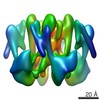 2526MC 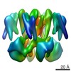 2527C 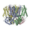 4chwC C: citing same article ( M: map data used to model this data |
|---|---|
| Similar structure data | |
| EM raw data | EMPIAR-10006 (Title: 2D crystal images of the potassium channel MloK1 with and without cAMP ligand Data size: 11.6 Data #1: Potassium channel MloK1 with cAMP ligand [micrographs - single frame] Data #2: Potassium channel MloK1 without cAMP ligand [micrographs - single frame]) |
-Links
 PDBj
PDBj


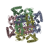
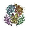
- Assembly
| Deposited unit | 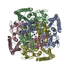
| ||||||||
|---|---|---|---|---|---|---|---|---|---|
| 1 |
| ||||||||
| Unit cell |
|
-Components
| #1: Protein | Mass: 38595.164 Da / Num. of mol.: 4 Source method: isolated from a genetically manipulated source Details: CAMP PRESENT IN BUFFER / Source: (gene. exp.) MESORHIZOBIUM LOTI (bacteria) / Plasmid: PASK90 / Production host: #2: Chemical |   Mass: 39.098 Da / Num. of mol.: 2 / Source method: obtained synthetically / Formula: K Mass: 39.098 Da / Num. of mol.: 2 / Source method: obtained synthetically / Formula: K |
|---|
-Experimental details
-Experiment
| Experiment | Method: ELECTRON CRYSTALLOGRAPHY |
|---|---|
| EM experiment | Aggregation state: 2D ARRAY / 3D reconstruction method: electron crystallography |
- Sample preparation
| Component | Name: MLOK1 WITH CAMP / Type: COMPLEX |
|---|---|
| Buffer solution | Name: 20 MM KCL, 1 MM BACL2, 1 MM EDTA, 20 MM TRIS, 0.2 MM CAMP pH: 7.6 Details: 20 MM KCL, 1 MM BACL2, 1 MM EDTA, 20 MM TRIS, 0.2 MM CAMP |
| Specimen | Conc.: 1.3 mg/ml / Embedding applied: NO / Shadowing applied: NO / Staining applied: NO / Vitrification applied: YES |
| Specimen support | Details: HOLEY CARBON |
| Vitrification | Instrument: FEI VITROBOT MARK IV / Cryogen name: ETHANE |
-Data collection
| Microscopy | Model: FEI/PHILIPS CM200FEG / Date: Mar 1, 2012 |
|---|---|
| Electron gun | Electron source:  FIELD EMISSION GUN / Accelerating voltage: 200 kV / Illumination mode: FLOOD BEAM FIELD EMISSION GUN / Accelerating voltage: 200 kV / Illumination mode: FLOOD BEAM |
| Electron lens | Mode: BRIGHT FIELD / Nominal magnification: 50000 X / Nominal defocus max: 3077 nm / Nominal defocus min: 655 nm / Cs: 2 mm |
| Specimen holder | Temperature: 85 K / Tilt angle max: 46 ° / Tilt angle min: 0 ° |
| Image recording | Electron dose: 5 e/Å2 / Film or detector model: KODAK SO-163 FILM |
| Image scans | Num. digital images: 78 |
| Radiation | Scattering type: electron |
| Radiation wavelength | Relative weight: 1 |
- Processing
| 3D reconstruction | Resolution: 7 Å / Resolution method: OTHER / Symmetry type: 2D CRYSTAL | ||||||||||||
|---|---|---|---|---|---|---|---|---|---|---|---|---|---|
| Refinement | Highest resolution: 7 Å | ||||||||||||
| Refinement step | Cycle: LAST / Highest resolution: 7 Å
|
