 Movie
Movie Controller

[English] 日本語
 Yorodumi
- PDB-4chm: Structure of Inner Membrane Complex (IMC) Sub-compartment Protein... -
+ Open data

- Basic information
| Entry | Database: PDB / ID: 4chm | ||||||
|---|---|---|---|---|---|---|---|
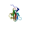
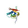
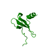
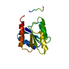
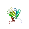
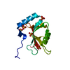
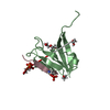
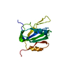
| Title | Structure of Inner Membrane Complex (IMC) Sub-compartment Protein 1 (ISP1) from Toxoplasma gondii | ||||||
 Components Components | IMC SUB-COMPARTMENT PROTEIN ISP1 | ||||||
 Keywords Keywords | CELL CYCLE / CELL DIVISION / ENDODYOGENY / PLECKSTRIN HOMOLOGY (PH) FOLD | ||||||
| Function / homology | ISP1 C-terminal / ISP1 C-terminal / Pleckstrin-homology domain (PH domain)/Phosphotyrosine-binding domain (PTB) / PH-domain like / PH-like domain superfamily / Roll / Mainly Beta / IMC sub-compartment protein ISP1 / IMC sub-compartment protein ISP1 Function and homology information Function and homology information | ||||||
| Biological species |  | ||||||
| Method |  X-RAY DIFFRACTION / SAD / Resolution: 2.1 Å X-RAY DIFFRACTION / SAD / Resolution: 2.1 Å | ||||||
 Authors Authors | Tonkin, M.L. / Beck, J.R. / Bradley, P.J. / Boulanger, M.J. | ||||||
 Citation Citation | Journal: J.Biol.Chem. / Year: 2014 Title: The Inner Membrane Complex Sub-Compartment Proteins Critical for Replication of the Apicomplexan Parasite Toxoplasma Gondii Adopt a Pleckstrin Homology Fold Authors: Tonkin, M.L. / Beck, J.R. / Bradley, P.J. / Boulanger, M.J. | ||||||
| History |
|
- Structure visualization
| Structure viewer | Molecule: MolmilJmol/JSmol |
|---|
- Downloads & links
-Download
| PDBx/mmCIF format | 4chm.cif.gz | 112.7 KB | Display | PDBx/mmCIF format |
|---|---|---|---|---|
| PDB format | pdb4chm.ent.gz | 88.3 KB | Display | PDB format |
| PDBx/mmJSON format | 4chm.json.gz | Tree view | PDBx/mmJSON format | |
| Others |  Other downloads Other downloads |
-Validation report
| Summary document | 4chm_validation.pdf.gz | 462.3 KB | Display | wwPDB validaton report |
|---|---|---|---|---|
| Full document | 4chm_full_validation.pdf.gz | 465.9 KB | Display | |
| Data in XML | 4chm_validation.xml.gz | 26.3 KB | Display | |
| Data in CIF | 4chm_validation.cif.gz | 35.3 KB | Display | |
| Arichive directory | https://data.pdbj.org/pub/pdb/validation_reports/ch/4chmftp://data.pdbj.org/pub/pdb/validation_reports/ch/4chm | HTTPS FTP |
-Related structure data

-Links
 PDBj
PDBj- Assembly
| Deposited unit | 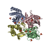
| ||||||||
|---|---|---|---|---|---|---|---|---|---|
| 1 | 
| ||||||||
| 2 | 
| ||||||||
| 3 | 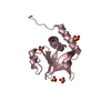
| ||||||||
| 4 | 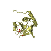
| ||||||||
| Unit cell |
|
-Components
| #1: Protein | Mass: 13844.036 Da / Num. of mol.: 4 / Fragment: PH FOLD CORE DOMAIN, RESIDUES 58-176 Source method: isolated from a genetically manipulated source Source: (gene. exp.)  #2: Chemical | ChemComp-SO4 /   Mass: 96.063 Da / Num. of mol.: 14 / Source method: obtained synthetically / Formula: SO4 Mass: 96.063 Da / Num. of mol.: 14 / Source method: obtained synthetically / Formula: SO4#3: Water | ChemComp-HOH / |  Mass: 18.015 Da / Num. of mol.: 272 / Source method: isolated from a natural source / Formula: H2O Mass: 18.015 Da / Num. of mol.: 272 / Source method: isolated from a natural source / Formula: H2OHas protein modification | Y | |
|---|
-Experimental details
-Experiment
| Experiment | Method: X-RAY DIFFRACTION |
|---|
- Sample preparation
| Crystal | Density Matthews: 2.44 Å3/Da / Density % sol: 49.56 % / Description: NONE |
|---|---|
| Crystal grow | Details: 0.1 M BIS-TRIS PH 5.5, 25% PEG 3350 |
-Data collection
| Diffraction | Mean temperature: 100 K |
|---|---|
| Diffraction source | Source: ROTATING ANODE / Type: RIGAKU MICROMAX-002 / Wavelength: 1.542 |
| Detector | Type: RIGAKU R-AXIS IV / Detector: IMAGE PLATE / Details: OSMIC BLUE |
| Radiation | Protocol: SINGLE WAVELENGTH / Monochromatic (M) / Laue (L): M / Scattering type: x-ray |
| Radiation wavelength | Wavelength: 1.542 Å / Relative weight: 1 |
| Reflection | Resolution: 2.1→29.09 Å / Num. obs: 33884 / % possible obs: 99.9 % / Observed criterion σ(I): 2 / Redundancy: 4.7 % / Rmerge(I) obs: 0.07 / Net I/σ(I): 16.3 |
| Reflection shell | Resolution: 2.1→2.21 Å / Redundancy: 4.8 % / Rmerge(I) obs: 0.38 / Mean I/σ(I) obs: 4.5 / % possible all: 100 |
- Processing
| Software |
| ||||||||||||||||||||||||||||||||||||||||||||||||||||||||||||||||||||||||||||||||||||||||||||||||||||||||||||||||||||||||||||||||||||||||||||||||||||||||||||||||||||||||||||||||||||||
|---|---|---|---|---|---|---|---|---|---|---|---|---|---|---|---|---|---|---|---|---|---|---|---|---|---|---|---|---|---|---|---|---|---|---|---|---|---|---|---|---|---|---|---|---|---|---|---|---|---|---|---|---|---|---|---|---|---|---|---|---|---|---|---|---|---|---|---|---|---|---|---|---|---|---|---|---|---|---|---|---|---|---|---|---|---|---|---|---|---|---|---|---|---|---|---|---|---|---|---|---|---|---|---|---|---|---|---|---|---|---|---|---|---|---|---|---|---|---|---|---|---|---|---|---|---|---|---|---|---|---|---|---|---|---|---|---|---|---|---|---|---|---|---|---|---|---|---|---|---|---|---|---|---|---|---|---|---|---|---|---|---|---|---|---|---|---|---|---|---|---|---|---|---|---|---|---|---|---|---|---|---|---|---|
| Refinement | Method to determine structure: SAD Starting model: NONE Resolution: 2.1→28.15 Å / Cor.coef. Fo:Fc: 0.944 / Cor.coef. Fo:Fc free: 0.916 / SU B: 5.2 / SU ML: 0.139 / Cross valid method: THROUGHOUT / ESU R: 0.229 / ESU R Free: 0.198 / Stereochemistry target values: MAXIMUM LIKELIHOOD / Details: HYDROGENS HAVE BEEN ADDED IN THE RIDING POSITIONS.
| ||||||||||||||||||||||||||||||||||||||||||||||||||||||||||||||||||||||||||||||||||||||||||||||||||||||||||||||||||||||||||||||||||||||||||||||||||||||||||||||||||||||||||||||||||||||
| Solvent computation | Ion probe radii: 0.8 Å / Shrinkage radii: 0.8 Å / VDW probe radii: 1.2 Å / Solvent model: MASK | ||||||||||||||||||||||||||||||||||||||||||||||||||||||||||||||||||||||||||||||||||||||||||||||||||||||||||||||||||||||||||||||||||||||||||||||||||||||||||||||||||||||||||||||||||||||
| Displacement parameters | Biso mean: 28.985 Å2
| ||||||||||||||||||||||||||||||||||||||||||||||||||||||||||||||||||||||||||||||||||||||||||||||||||||||||||||||||||||||||||||||||||||||||||||||||||||||||||||||||||||||||||||||||||||||
| Refinement step | Cycle: LAST / Resolution: 2.1→28.15 Å
| ||||||||||||||||||||||||||||||||||||||||||||||||||||||||||||||||||||||||||||||||||||||||||||||||||||||||||||||||||||||||||||||||||||||||||||||||||||||||||||||||||||||||||||||||||||||
| Refine LS restraints |
|