 Movie
Movie Controller

[English] 日本語
 Yorodumi
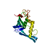
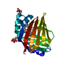
- PDB-4chd: Crystal structure of the '627' domain of the PB2 subunit of Thogo... -
+ Open data

- Basic information
| Entry | Database: PDB / ID: 4chd | ||||||
|---|---|---|---|---|---|---|---|
| Title | Crystal structure of the '627' domain of the PB2 subunit of Thogoto virus polymerase | ||||||
 Components Components | POLYMERASE ACIDIC PROTEIN | ||||||
 Keywords Keywords | VIRAL PROTEIN | ||||||
| Function / homology |  Function and homology information Function and homology informationcap snatching / 7-methylguanosine mRNA capping / virion component / host cell nucleus Similarity search - Function | ||||||
| Biological species |  THOGOTO VIRUS THOGOTO VIRUS | ||||||
| Method |  X-RAY DIFFRACTION / SYNCHROTRON / SAD / Resolution: 2.4 Å X-RAY DIFFRACTION / SYNCHROTRON / SAD / Resolution: 2.4 Å | ||||||
 Authors Authors | Guilligay, D. / Kadlec, J. / Crepin, T. / Lunardi, T. / Bouvier, D. / Kochs, G. / Ruigrok, R.W.H. / Cusack, S. | ||||||
 Citation Citation | Journal: Plos One / Year: 2014 Title: Comparative Structural and Functional Analysis of Orthomyxovirus Polymerase CAP-Snatching Domains. Authors: Guilligay, D. / Kadlec, J. / Crepin, T. / Lunardi, T. / Bouvier, D. / Kochs, G. / Ruigrok, R.W.H. / Cusack, S. | ||||||
| History |
|
- Structure visualization
| Structure viewer | Molecule: MolmilJmol/JSmol |
|---|
- Downloads & links
-Download
| PDBx/mmCIF format | 4chd.cif.gz | 68.1 KB | Display | PDBx/mmCIF format |
|---|---|---|---|---|
| PDB format | pdb4chd.ent.gz | 50.7 KB | Display | PDB format |
| PDBx/mmJSON format | 4chd.json.gz | Tree view | PDBx/mmJSON format | |
| Others |  Other downloads Other downloads |
-Validation report
| Arichive directory | https://data.pdbj.org/pub/pdb/validation_reports/ch/4chdftp://data.pdbj.org/pub/pdb/validation_reports/ch/4chd | HTTPS FTP |
|---|
-Related structure data


-Links
 PDBj
PDBj- Assembly
| Deposited unit | 
| ||||||||
|---|---|---|---|---|---|---|---|---|---|
| 1 |
| ||||||||
| Unit cell |
|
-Components
| #1: Protein | Mass: 18304.828 Da / Num. of mol.: 1 / Fragment: CENTRAL '627' DOMAIN, RESIDUES 543-701 Source method: isolated from a genetically manipulated source Source: (gene. exp.) THOGOTO VIRUS / Production host:  |
|---|---|
| #2: Water | ChemComp-HOH /  Mass: 18.015 Da / Num. of mol.: 17 / Source method: isolated from a natural source / Formula: H2O Mass: 18.015 Da / Num. of mol.: 17 / Source method: isolated from a natural source / Formula: H2O |
| Sequence details | EXTRA GAM AT N-TERMINUS AFTER HIS-TAG CLEAVAGE |
-Experimental details
-Experiment
| Experiment | Method: X-RAY DIFFRACTION / Number of used crystals: 1 |
|---|
- Sample preparation
| Crystal | Density Matthews: 2.2 Å3/Da / Density % sol: 44.1 % / Description: SOLVED WITH SINGLE SITE MERCURY DERIVATIVE |
|---|---|
| Crystal grow | pH: 7 Details: THE PROTEIN WAS CONCENTRATED TO 2.3 MG/ML IN 250 MM NACL, 50 MM TRIS PH 8.0 AND 2 MM DTT AND CRYSTALS WERE GROWN IN 0.1 M HEPES PH 7.0, 0.2 M AMMONIUM SULPHATE AND 22% PEG3350 |
-Data collection
| Diffraction | Mean temperature: 100 K |
|---|---|
| Diffraction source | Source: SYNCHROTRON / Site: ESRF  / Beamline: ID23-2 / Wavelength: 0.8726 / Beamline: ID23-2 / Wavelength: 0.8726 |
| Detector | Type: MARMOSAIC 225 mm CCD / Detector: CCD / Date: Jul 10, 2008 |
| Radiation | Protocol: SINGLE WAVELENGTH / Monochromatic (M) / Laue (L): M / Scattering type: x-ray |
| Radiation wavelength | Wavelength: 0.8726 Å / Relative weight: 1 |
| Reflection | Resolution: 2.4→50 Å / Num. obs: 6604 / % possible obs: 97.4 % / Observed criterion σ(I): 0 / Redundancy: 3.95 % / Rmerge(I) obs: 0.08 / Net I/σ(I): 11.1 |
| Reflection shell | Resolution: 2.4→2.5 Å / Redundancy: 4.01 % / Rmerge(I) obs: 0.56 / Mean I/σ(I) obs: 2.6 / % possible all: 98.7 |
- Processing
| Software |
| ||||||||||||||||||||||||||||||||||||||||||||||||||||||||||||||||||||||||||||||||||||||||||||||||||||||||||||||||||||||||||||||||||||||||||||||||||||||||||||||||||||||||||||||||||||||
|---|---|---|---|---|---|---|---|---|---|---|---|---|---|---|---|---|---|---|---|---|---|---|---|---|---|---|---|---|---|---|---|---|---|---|---|---|---|---|---|---|---|---|---|---|---|---|---|---|---|---|---|---|---|---|---|---|---|---|---|---|---|---|---|---|---|---|---|---|---|---|---|---|---|---|---|---|---|---|---|---|---|---|---|---|---|---|---|---|---|---|---|---|---|---|---|---|---|---|---|---|---|---|---|---|---|---|---|---|---|---|---|---|---|---|---|---|---|---|---|---|---|---|---|---|---|---|---|---|---|---|---|---|---|---|---|---|---|---|---|---|---|---|---|---|---|---|---|---|---|---|---|---|---|---|---|---|---|---|---|---|---|---|---|---|---|---|---|---|---|---|---|---|---|---|---|---|---|---|---|---|---|---|---|
| Refinement | Method to determine structure: SAD Starting model: NONE Resolution: 2.4→49.27 Å / Cor.coef. Fo:Fc: 0.946 / Cor.coef. Fo:Fc free: 0.926 / SU B: 16.159 / SU ML: 0.175 / Cross valid method: THROUGHOUT / ESU R: 0.384 / ESU R Free: 0.245 / Stereochemistry target values: MAXIMUM LIKELIHOOD Details: HYDROGENS HAVE BEEN ADDED IN THE RIDING POSITIONS. U VALUES WITH TLS ADDED
| ||||||||||||||||||||||||||||||||||||||||||||||||||||||||||||||||||||||||||||||||||||||||||||||||||||||||||||||||||||||||||||||||||||||||||||||||||||||||||||||||||||||||||||||||||||||
| Solvent computation | Ion probe radii: 0.8 Å / Shrinkage radii: 0.8 Å / VDW probe radii: 1.2 Å / Solvent model: MASK | ||||||||||||||||||||||||||||||||||||||||||||||||||||||||||||||||||||||||||||||||||||||||||||||||||||||||||||||||||||||||||||||||||||||||||||||||||||||||||||||||||||||||||||||||||||||
| Displacement parameters | Biso mean: 50.362 Å2
| ||||||||||||||||||||||||||||||||||||||||||||||||||||||||||||||||||||||||||||||||||||||||||||||||||||||||||||||||||||||||||||||||||||||||||||||||||||||||||||||||||||||||||||||||||||||
| Refinement step | Cycle: LAST / Resolution: 2.4→49.27 Å
| ||||||||||||||||||||||||||||||||||||||||||||||||||||||||||||||||||||||||||||||||||||||||||||||||||||||||||||||||||||||||||||||||||||||||||||||||||||||||||||||||||||||||||||||||||||||
| Refine LS restraints |
|