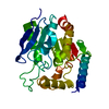
 Movie
Movie Controller

[English] 日本語
 Yorodumi
- PDB-4cg3: Structural and functional studies on a thermostable polyethylene ... -
+ Open data

- Basic information
| Entry | Database: PDB / ID: 4cg3 | ||||||
|---|---|---|---|---|---|---|---|
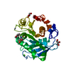
| Title | Structural and functional studies on a thermostable polyethylene therephtalate degrading hydrolase from Thermobifida fusca | ||||||
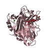
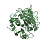
 Components Components | CUTINASE | ||||||
 Keywords Keywords | HYDROLASE / PET DEGRADATION / ALPHA-BETA- FOLD | ||||||
| Function / homology |  Function and homology information Function and homology informationpoly(ethylene terephthalate) hydrolase / cutinase / cutinase activity / periplasmic space / extracellular region Similarity search - Function | ||||||
| Biological species |   THERMOBIFIDA FUSCA (bacteria) THERMOBIFIDA FUSCA (bacteria) | ||||||
| Method |  X-RAY DIFFRACTION / MOLECULAR REPLACEMENT / Resolution: 1.55 Å X-RAY DIFFRACTION / MOLECULAR REPLACEMENT / Resolution: 1.55 Å | ||||||
 Authors Authors | Roth, C. / Wei, R. / Oeser, T. / Then, J. / Foellner, C. / Zimmermann, W. / Straeter, N. | ||||||
 Citation Citation | Journal: Appl.Microbiol.Biotechnol. / Year: 2014 Title: Structural and Functional Studies on a Thermostable Polyethylene Terephthalate Degrading Hydrolase from Thermobifida Fusca. Authors: Roth, C. / Wei, R. / Oeser, T. / Then, J. / Follner, C. / Zimmermann, W. / Strater, N. | ||||||
| History |
|
- Structure visualization
| Structure viewer | Molecule: MolmilJmol/JSmol |
|---|
- Downloads & links
-Download
| PDBx/mmCIF format | 4cg3.cif.gz | 13.7 MB | Display | PDBx/mmCIF format |
|---|---|---|---|---|
| PDB format | pdb4cg3.ent.gz | 11.7 MB | Display | PDB format |
| PDBx/mmJSON format | 4cg3.json.gz | Tree view | PDBx/mmJSON format | |
| Others |  Other downloads Other downloads |
-Validation report
| Arichive directory | https://data.pdbj.org/pub/pdb/validation_reports/cg/4cg3ftp://data.pdbj.org/pub/pdb/validation_reports/cg/4cg3 | HTTPS FTP |
|---|
-Related structure data
| Related structure data |  4cg1C 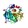 4cg2C  1jfrS C: citing same article ( S: Starting model for refinement |
|---|---|
| Similar structure data |


-Links
 PDBj
PDBj
- Assembly
| Deposited unit | 
| ||||||||
|---|---|---|---|---|---|---|---|---|---|
| 1 |
| ||||||||
| Unit cell |
| ||||||||
| Number of models | 77 |
-Components
| #1: Protein | Mass: 33789.965 Da / Num. of mol.: 1 Source method: isolated from a genetically manipulated source Source: (gene. exp.) THERMOBIFIDA FUSCA (bacteria) / Strain: KW3 / Production host: |
|---|---|
| #2: Chemical | ChemComp-SO4 /   Mass: 96.063 Da / Num. of mol.: 1 / Source method: obtained synthetically / Formula: SO4 Mass: 96.063 Da / Num. of mol.: 1 / Source method: obtained synthetically / Formula: SO4 |
| #3: Water | ChemComp-HOH /  Mass: 18.015 Da / Num. of mol.: 99 / Source method: isolated from a natural source / Formula: H2O Mass: 18.015 Da / Num. of mol.: 99 / Source method: isolated from a natural source / Formula: H2O |
| Has protein modification | Y |
-Experimental details
-Experiment
| Experiment | Method: X-RAY DIFFRACTION |
|---|
- Sample preparation
| Crystal | Density Matthews: 2.2 Å3/Da / Density % sol: 45.2 % / Description: NONE |
|---|
-Data collection
| Diffraction | Mean temperature: 100 K |
|---|---|
| Diffraction source | Source: ROTATING ANODE / Type: BRUKER AXS MICROSTAR / Wavelength: 1.541 |
| Detector | Type: MARRESEARCH |
| Radiation | Protocol: SINGLE WAVELENGTH / Monochromatic (M) / Laue (L): M / Scattering type: x-ray |
| Radiation wavelength | Wavelength: 1.541 Å / Relative weight: 1 |
| Reflection | Resolution: 1.55→23 Å / Num. obs: 36710 / % possible obs: 100 % / Observed criterion σ(I): 2.55 / Redundancy: 3.8 % / Biso Wilson estimate: 16 Å2 / Rmerge(I) obs: 0.1 / Net I/σ(I): 7.6 |
| Reflection shell | Resolution: 1.55→1.58 Å / Redundancy: 3.7 % / Rmerge(I) obs: 0.59 / Mean I/σ(I) obs: 2.4 / % possible all: 100 |
- Processing
| Software |
| ||||||||||||||||||||||||||||||||||||||||||||||||||||||||||||||||||||||||||||||||||||||||||||||||||||||||||||||||||||||||||||||||||||||||||||||||||||||||||||||||||||||||
|---|---|---|---|---|---|---|---|---|---|---|---|---|---|---|---|---|---|---|---|---|---|---|---|---|---|---|---|---|---|---|---|---|---|---|---|---|---|---|---|---|---|---|---|---|---|---|---|---|---|---|---|---|---|---|---|---|---|---|---|---|---|---|---|---|---|---|---|---|---|---|---|---|---|---|---|---|---|---|---|---|---|---|---|---|---|---|---|---|---|---|---|---|---|---|---|---|---|---|---|---|---|---|---|---|---|---|---|---|---|---|---|---|---|---|---|---|---|---|---|---|---|---|---|---|---|---|---|---|---|---|---|---|---|---|---|---|---|---|---|---|---|---|---|---|---|---|---|---|---|---|---|---|---|---|---|---|---|---|---|---|---|---|---|---|---|---|---|---|---|
| Refinement | Method to determine structure: MOLECULAR REPLACEMENT Starting model: PDB ENTRY 1JFR Resolution: 1.55→23.122 Å / SU ML: 0.11 / σ(F): 1.24 / Phase error: 13.59 / Stereochemistry target values: ML / Details: REFINED USING PHENIX.ENSEMBLE REFINEMENT
| ||||||||||||||||||||||||||||||||||||||||||||||||||||||||||||||||||||||||||||||||||||||||||||||||||||||||||||||||||||||||||||||||||||||||||||||||||||||||||||||||||||||||
| Solvent computation | Shrinkage radii: 0.8 Å / VDW probe radii: 1 Å / Solvent model: FLAT BULK SOLVENT MODEL | ||||||||||||||||||||||||||||||||||||||||||||||||||||||||||||||||||||||||||||||||||||||||||||||||||||||||||||||||||||||||||||||||||||||||||||||||||||||||||||||||||||||||
| Displacement parameters | Biso mean: 17.1 Å2 | ||||||||||||||||||||||||||||||||||||||||||||||||||||||||||||||||||||||||||||||||||||||||||||||||||||||||||||||||||||||||||||||||||||||||||||||||||||||||||||||||||||||||
| Refinement step | Cycle: LAST / Resolution: 1.55→23.122 Å
| ||||||||||||||||||||||||||||||||||||||||||||||||||||||||||||||||||||||||||||||||||||||||||||||||||||||||||||||||||||||||||||||||||||||||||||||||||||||||||||||||||||||||
| LS refinement shell |
|
