 Movie
Movie Controller


+ Open data

- Basic information
| Entry | Database: PDB / ID: 4ceh | ||||||
|---|---|---|---|---|---|---|---|
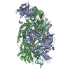
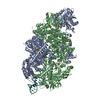
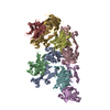
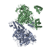
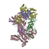
| Title | Crystal structure of AddAB with a forked DNA substrate | ||||||
 Components Components |
| ||||||
 Keywords Keywords | HYDROLASE/DNA / HYDROLASE-DNA COMPLEX / HELICASE-NUCLEASE / BACTERIAL PROTEINS / BINDING SITES / DNA BREAKS / DOUBLE-STRANDED / DNA HELICASES / DNA REPAIR / DNA- BINDING PROTEINS / EXODEOXYRIBONUCLEASE V / EXODEOXYRIBONUCLEASES / HOMOLOGOUS RECOMBINATION | ||||||
| Function / homology |  Function and homology information Function and homology informationDNA helicase complex / 5'-3' exonuclease activity / recombinational repair / DNA 3'-5' helicase / 3'-5' DNA helicase activity / 3'-5' exonuclease activity / helicase activity / double-strand break repair via homologous recombination / 4 iron, 4 sulfur cluster binding / double-stranded DNA binding ...DNA helicase complex / 5'-3' exonuclease activity / recombinational repair / DNA 3'-5' helicase / 3'-5' DNA helicase activity / 3'-5' exonuclease activity / helicase activity / double-strand break repair via homologous recombination / 4 iron, 4 sulfur cluster binding / double-stranded DNA binding / DNA recombination / Hydrolases; Acting on ester bonds / ATP hydrolysis activity / ATP binding / metal ion binding / cytosol Similarity search - Function | ||||||
| Biological species |  SYNTHETIC CONSTRUCT (others) | ||||||
| Method |  X-RAY DIFFRACTION / SYNCHROTRON / MOLECULAR REPLACEMENT / Resolution: 3.24 Å X-RAY DIFFRACTION / SYNCHROTRON / MOLECULAR REPLACEMENT / Resolution: 3.24 Å | ||||||
 Authors Authors | Krajewski, W.W. / Wilkinson, M. / Fu, X. / Cronin, N.B. / Wigley, D. | ||||||
 Citation Citation | Journal: Nature / Year: 2014 Title: Structural Basis for Translocation by Addab Helicase-Nuclease and its Arrest at Chi Sites. Authors: Krajewski, W.W. / Fu, X. / Wilkinson, M. / Cronin, N.B. / Dillingham, M.S. / Wigley, D.B. | ||||||
| History |
|
- Structure visualization
| Structure viewer | Molecule: MolmilJmol/JSmol |
|---|
- Downloads & links
-Download
| PDBx/mmCIF format | 4ceh.cif.gz | 497.1 KB | Display | PDBx/mmCIF format |
|---|---|---|---|---|
| PDB format | pdb4ceh.ent.gz | 394.4 KB | Display | PDB format |
| PDBx/mmJSON format | 4ceh.json.gz | Tree view | PDBx/mmJSON format | |
| Others |  Other downloads Other downloads |
-Validation report
| Arichive directory | https://data.pdbj.org/pub/pdb/validation_reports/ce/4cehftp://data.pdbj.org/pub/pdb/validation_reports/ce/4ceh | HTTPS FTP |
|---|
-Related structure data
| Related structure data |  4ceiC  4cejC 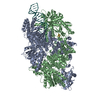 3u44S C: citing same article ( S: Starting model for refinement |
|---|---|
| Similar structure data |


-Links
 PDBj
PDBj


- Assembly
| Deposited unit | 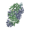
| ||||||||
|---|---|---|---|---|---|---|---|---|---|
| 1 |
| ||||||||
| Unit cell |
|
-Components
| #1: Protein | Mass: 141218.844 Da / Num. of mol.: 1 / Mutation: YES Source method: isolated from a genetically manipulated source Source: (gene. exp.) Plasmid: PCOLADUET-1 / Production host: References: UniProt: P23478, Hydrolases; Acting on ester bonds, DNA helicase |
|---|---|
| #2: Protein | Mass: 134769.406 Da / Num. of mol.: 1 / Mutation: YES Source method: isolated from a genetically manipulated source Source: (gene. exp.) Plasmid: PCOLADUET-1 / Production host: References: UniProt: P23477, Hydrolases; Acting on ester bonds, DNA helicase |
| #3: DNA chain | Mass: 19956.744 Da / Num. of mol.: 1 / Source method: obtained synthetically / Details: SYNTHETIC CONSTRUCT / Source: (synth.) SYNTHETIC CONSTRUCT (others) |
| #4: Chemical | ChemComp-SF4 /   Mass: 351.640 Da / Num. of mol.: 1 / Source method: obtained synthetically / Formula: Fe4S4 Mass: 351.640 Da / Num. of mol.: 1 / Source method: obtained synthetically / Formula: Fe4S4 |
-Experimental details
-Experiment
| Experiment | Method: X-RAY DIFFRACTION / Number of used crystals: 1 |
|---|
- Sample preparation
| Crystal | Density Matthews: 2.99 Å3/Da / Density % sol: 59 % / Description: NONE |
|---|---|
| Crystal grow | Temperature: 285 K / Method: vapor diffusion, hanging drop / pH: 7.5 Details: 0.1M TRIS-HCL PH 7.5, 15% PEG 4000, 0.8M SODIUM FORMATE, VAPOR DIFFUSION, HANGING DROP, 285K |
-Data collection
| Diffraction | Mean temperature: 100 K |
|---|---|
| Diffraction source | Source: SYNCHROTRON / Site: Diamond  / Beamline: I02 / Wavelength: 0.97949 / Beamline: I02 / Wavelength: 0.97949 |
| Detector | Type: DECTRIS PILATUS 6M / Detector: PIXEL / Date: Oct 8, 2012 |
| Radiation | Protocol: SINGLE WAVELENGTH / Monochromatic (M) / Laue (L): M / Scattering type: x-ray |
| Radiation wavelength | Wavelength: 0.97949 Å / Relative weight: 1 |
| Reflection | Resolution: 3.24→39.52 Å / Num. obs: 47940 / % possible obs: 98.5 % / Observed criterion σ(I): 2 / Redundancy: 2.9 % / Rmerge(I) obs: 0.05 / Net I/σ(I): 13.2 |
| Reflection shell | Resolution: 3.24→3.35 Å / Redundancy: 2.9 % / Rmerge(I) obs: 0.33 / Mean I/σ(I) obs: 2.3 / % possible all: 97.7 |
- Processing
| Software |
| ||||||||||||||||||||||||||||||||||||||||||||||||||||||||||||||||||||||||||||||||||||||||||||||||||||||||||||||||||||||||||||||
|---|---|---|---|---|---|---|---|---|---|---|---|---|---|---|---|---|---|---|---|---|---|---|---|---|---|---|---|---|---|---|---|---|---|---|---|---|---|---|---|---|---|---|---|---|---|---|---|---|---|---|---|---|---|---|---|---|---|---|---|---|---|---|---|---|---|---|---|---|---|---|---|---|---|---|---|---|---|---|---|---|---|---|---|---|---|---|---|---|---|---|---|---|---|---|---|---|---|---|---|---|---|---|---|---|---|---|---|---|---|---|---|---|---|---|---|---|---|---|---|---|---|---|---|---|---|---|---|
| Refinement | Method to determine structure: MOLECULAR REPLACEMENT Starting model: PDB ENTRY 3U44 Resolution: 3.24→29.939 Å / SU ML: 0.42 / σ(F): 1.97 / Phase error: 32.34 / Stereochemistry target values: ML
| ||||||||||||||||||||||||||||||||||||||||||||||||||||||||||||||||||||||||||||||||||||||||||||||||||||||||||||||||||||||||||||||
| Solvent computation | Shrinkage radii: 0.9 Å / VDW probe radii: 1.11 Å / Solvent model: FLAT BULK SOLVENT MODEL | ||||||||||||||||||||||||||||||||||||||||||||||||||||||||||||||||||||||||||||||||||||||||||||||||||||||||||||||||||||||||||||||
| Refinement step | Cycle: LAST / Resolution: 3.24→29.939 Å
| ||||||||||||||||||||||||||||||||||||||||||||||||||||||||||||||||||||||||||||||||||||||||||||||||||||||||||||||||||||||||||||||
| Refine LS restraints |
| ||||||||||||||||||||||||||||||||||||||||||||||||||||||||||||||||||||||||||||||||||||||||||||||||||||||||||||||||||||||||||||||
| LS refinement shell |
|
