 Movie
Movie Controller

[English] 日本語
 Yorodumi
- PDB-4ce8: Perdeuterated Pseudomonas aeruginosa Lectin II complex with hydro... -
+ Open data

- Basic information
| Entry | Database: PDB / ID: 4ce8 | |||||||||
|---|---|---|---|---|---|---|---|---|---|---|
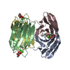
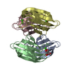
| Title | Perdeuterated Pseudomonas aeruginosa Lectin II complex with hydrogenated L-Fucose and Calcium | |||||||||
 Components Components | FUCOSE-BINDING LECTIN PA-IIL | |||||||||
 Keywords Keywords | SUGAR BINDING PROTEIN / PERDEUTERATED | |||||||||
| Function / homology |  Function and homology information Function and homology informationsingle-species biofilm formation / carbohydrate binding / metal ion binding Similarity search - Function | |||||||||
| Biological species |   PSEUDOMONAS AERUGINOSA (bacteria) PSEUDOMONAS AERUGINOSA (bacteria) | |||||||||
| Method |  X-RAY DIFFRACTION / SYNCHROTRON / MOLECULAR REPLACEMENT / Resolution: 0.9 Å X-RAY DIFFRACTION / SYNCHROTRON / MOLECULAR REPLACEMENT / Resolution: 0.9 Å | |||||||||
 Authors Authors | Cuypers, M.G. / Mitchell, E.P. / Mossou, E. / Pokorna, M. / Wimmerova, M. / Imberty, A. / Moulin, M. / Haertlein, M. / Forsyth, V.T. | |||||||||
 Citation Citation | Journal: To be Published Title: Perdeuterated Pseudomonas Aeruginosa Lectin II Complex with Hydrogenated L Fucose and Calcium Authors: Cuypers, M.G. / Mossou, E. / Mitchell, E.P. / Russi, S. / Pokorna, M. / Wimmerova, M. / Imberty, A. / Mcsweeney, S. / Haertlein, M. / Forsyth, V.T. | |||||||||
| History |
|
- Structure visualization
| Structure viewer | Molecule: MolmilJmol/JSmol |
|---|
- Downloads & links
-Download
| PDBx/mmCIF format | 4ce8.cif.gz | 306.9 KB | Display | PDBx/mmCIF format |
|---|---|---|---|---|
| PDB format | pdb4ce8.ent.gz | 252.5 KB | Display | PDB format |
| PDBx/mmJSON format | 4ce8.json.gz | Tree view | PDBx/mmJSON format | |
| Others |  Other downloads Other downloads |
-Validation report
| Arichive directory | https://data.pdbj.org/pub/pdb/validation_reports/ce/4ce8ftp://data.pdbj.org/pub/pdb/validation_reports/ce/4ce8 | HTTPS FTP |
|---|
-Related structure data
| Related structure data |  1uzvS S: Starting model for refinement |
|---|---|
| Similar structure data |
-Links
 PDBj
PDBj
- Assembly
| Deposited unit | 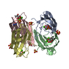
| ||||||||
|---|---|---|---|---|---|---|---|---|---|
| 1 |
| ||||||||
| Unit cell |
|
-Components
-Protein / Sugars , 2 types, 8 molecules ABCD
| #1: Protein | Mass: 11734.707 Da / Num. of mol.: 4 / Fragment: 2-115 Source method: isolated from a genetically manipulated source Details: PERDEUTERATED PROTEIN / Source: (gene. exp.) PSEUDOMONAS AERUGINOSA (bacteria) / Plasmid: PET28A / Production host: #3: Sugar | ChemComp-FUC /  Type: L-saccharide, alpha linking / Mass: 164.156 Da / Num. of mol.: 4 Type: L-saccharide, alpha linking / Mass: 164.156 Da / Num. of mol.: 4Source method: isolated from a genetically manipulated source Formula: C6H12O5 |
|---|
-Non-polymers , 4 types, 1007 molecules 


| #2: Chemical | ChemComp-CA /  Mass: 40.078 Da / Num. of mol.: 8 / Source method: obtained synthetically / Formula: Ca Mass: 40.078 Da / Num. of mol.: 8 / Source method: obtained synthetically / Formula: Ca#4: Chemical | ChemComp-SO4 /  Mass: 96.063 Da / Num. of mol.: 9 / Source method: obtained synthetically / Formula: SO4 Mass: 96.063 Da / Num. of mol.: 9 / Source method: obtained synthetically / Formula: SO4#5: Chemical | ChemComp-URE / |  Mass: 60.055 Da / Num. of mol.: 1 / Source method: obtained synthetically / Formula: CH4N2O Mass: 60.055 Da / Num. of mol.: 1 / Source method: obtained synthetically / Formula: CH4N2O#6: Water | ChemComp-HOH / | Mass: 18.015 Da / Num. of mol.: 989 / Source method: isolated from a natural source / Formula: H2O |
|---|
-Experimental details
-Experiment
| Experiment | Method: X-RAY DIFFRACTION / Number of used crystals: 1 |
|---|
- Sample preparation
| Crystal | Density Matthews: 2.21 Å3/Da / Density % sol: 44.28 % Description: NEEDED ONLY 2 CA ATOMS FROM 1UZV FOR ISOMORPHOUS REPLACEMENT WITH ACORN. |
|---|---|
| Crystal grow | Method: vapor diffusion, hanging drop Details: ALL SALTS EXCHANGED 3X AGAINST D2O. 0,1 M TRIS PD 8.5, 1.75M (ND4)2SO4, 2MM CACL2, 2MM MGCL2. 10 MG/ML PROTEIN PRE-INCUBATED IN 2MM CACL2 AND 250 MG/L L-FUCOSE. HANGING DROP METHOD. |
-Data collection
| Diffraction | Mean temperature: 100 K |
|---|---|
| Diffraction source | Source: SYNCHROTRON / Site: ESRF  / Beamline: ID23-1 / Wavelength: 0.8266 / Beamline: ID23-1 / Wavelength: 0.8266 |
| Detector | Type: ADSC CCD / Detector: CCD / Date: Jan 1, 2009 |
| Radiation | Protocol: SINGLE WAVELENGTH / Monochromatic (M) / Laue (L): M / Scattering type: x-ray |
| Radiation wavelength | Wavelength: 0.8266 Å / Relative weight: 1 |
| Reflection | Resolution: 0.9→43.55 Å / Num. obs: 291511 / % possible obs: 97.1 % / Observed criterion σ(I): 1.8 / Redundancy: 3 % / Biso Wilson estimate: 6.16 Å2 / Rmerge(I) obs: 0.08 / Net I/σ(I): 8.2 |
| Reflection shell | Highest resolution: 0.9 Å / Redundancy: 2.7 % / Rmerge(I) obs: 0.68 / Mean I/σ(I) obs: 1.8 / % possible all: 94.7 |
- Processing
| Software |
| |||||||||||||||||||||||||||||||||||||||||||||||||||||||||||||||||||||||||||||||||||||||||||||||||||||||||||||||||||||||||||||||||||||||||||||||||||||||||||||||||||||||||||||||||||||||||||||||||||||||||||||||||||||||||
|---|---|---|---|---|---|---|---|---|---|---|---|---|---|---|---|---|---|---|---|---|---|---|---|---|---|---|---|---|---|---|---|---|---|---|---|---|---|---|---|---|---|---|---|---|---|---|---|---|---|---|---|---|---|---|---|---|---|---|---|---|---|---|---|---|---|---|---|---|---|---|---|---|---|---|---|---|---|---|---|---|---|---|---|---|---|---|---|---|---|---|---|---|---|---|---|---|---|---|---|---|---|---|---|---|---|---|---|---|---|---|---|---|---|---|---|---|---|---|---|---|---|---|---|---|---|---|---|---|---|---|---|---|---|---|---|---|---|---|---|---|---|---|---|---|---|---|---|---|---|---|---|---|---|---|---|---|---|---|---|---|---|---|---|---|---|---|---|---|---|---|---|---|---|---|---|---|---|---|---|---|---|---|---|---|---|---|---|---|---|---|---|---|---|---|---|---|---|---|---|---|---|---|---|---|---|---|---|---|---|---|---|---|---|---|---|---|---|---|
| Refinement | Method to determine structure: MOLECULAR REPLACEMENT Starting model: PDB ENTRY 1UZV Resolution: 0.9→36.463 Å / SU ML: 0.07 / σ(F): 1.34 / Phase error: 13.29 / Stereochemistry target values: ML Details: UREA FOUND NEARBY ARGININES, PARTIAL DEGRADATION POSSIBLE INTO ORNITHINE.
| |||||||||||||||||||||||||||||||||||||||||||||||||||||||||||||||||||||||||||||||||||||||||||||||||||||||||||||||||||||||||||||||||||||||||||||||||||||||||||||||||||||||||||||||||||||||||||||||||||||||||||||||||||||||||
| Solvent computation | Shrinkage radii: 0.9 Å / VDW probe radii: 1.11 Å / Solvent model: FLAT BULK SOLVENT MODEL | |||||||||||||||||||||||||||||||||||||||||||||||||||||||||||||||||||||||||||||||||||||||||||||||||||||||||||||||||||||||||||||||||||||||||||||||||||||||||||||||||||||||||||||||||||||||||||||||||||||||||||||||||||||||||
| Displacement parameters | Biso mean: 4.23 Å2 | |||||||||||||||||||||||||||||||||||||||||||||||||||||||||||||||||||||||||||||||||||||||||||||||||||||||||||||||||||||||||||||||||||||||||||||||||||||||||||||||||||||||||||||||||||||||||||||||||||||||||||||||||||||||||
| Refinement step | Cycle: LAST / Resolution: 0.9→36.463 Å
| |||||||||||||||||||||||||||||||||||||||||||||||||||||||||||||||||||||||||||||||||||||||||||||||||||||||||||||||||||||||||||||||||||||||||||||||||||||||||||||||||||||||||||||||||||||||||||||||||||||||||||||||||||||||||
| Refine LS restraints |
| |||||||||||||||||||||||||||||||||||||||||||||||||||||||||||||||||||||||||||||||||||||||||||||||||||||||||||||||||||||||||||||||||||||||||||||||||||||||||||||||||||||||||||||||||||||||||||||||||||||||||||||||||||||||||
| LS refinement shell |
|
