 Movie
Movie Controller

[English] 日本語
 Yorodumi
- PDB-2bp6: crystal Structure of pseudomonas aeruginosa lectin (PA-IIL) compl... -
+ Open data

- Basic information
| Entry | Database: PDB / ID: 2bp6 | ||||||
|---|---|---|---|---|---|---|---|
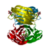
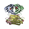
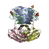
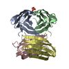
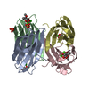
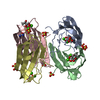
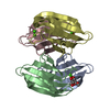
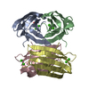
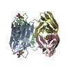
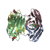
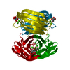
| Title | crystal Structure of pseudomonas aeruginosa lectin (PA-IIL) complexed with a-L-Galactopyranoside | ||||||
 Components Components | PSEUDOMONAS AERUGINOSA LECTIN II | ||||||
 Keywords Keywords | LECTIN / GALACTOSE / CALCIUM / PSEUDOMONAS AERUGINOSA | ||||||
| Function / homology |  Function and homology information Function and homology informationsingle-species biofilm formation / carbohydrate binding / metal ion binding Similarity search - Function | ||||||
| Biological species |   PSEUDOMONAS AERUGINOSA (bacteria) PSEUDOMONAS AERUGINOSA (bacteria) | ||||||
| Method |  X-RAY DIFFRACTION / SYNCHROTRON / MOLECULAR REPLACEMENT / Resolution: 1.5 Å X-RAY DIFFRACTION / SYNCHROTRON / MOLECULAR REPLACEMENT / Resolution: 1.5 Å | ||||||
 Authors Authors | Sabin, C.D. / Mitchell, E.P. / Wimmerova, M. / Imberty, A. | ||||||
 Citation Citation | Journal: FEBS Lett. / Year: 2006 Title: Binding of Different Monosaccharides by Lectin Pa-Iil from Pseudoman Aeruginosa: Thermodynamics Data Correlated with X-Ray Structures. Authors: Sabin, C.D. / Mitchell, E.P. / Pokarna, M. / Gautier, C. / Utille, J.-P. / Wimmerova, M. / Imberty, A. #1: Journal: Proteins: Struct.,Funct., Genet. / Year: 2005Title: High Affinity Fucose Binding of Pseudomonas Aeruginosa Lectin Pa-Iil: 1.0 A Resolution Crystal Structure of the Complex Combined with Thermodynamics and Computational Chemistry Approaches Authors: Mitchell, E. / Sabin, C.D. / Snajdrova, L. / Pokorna, M. / Perret, S. / Gautier, C. / Hofr, C. / Gilboa-Garber, N. / Koca, J. / Wimmerova, M. / Imberty, A. #2: Journal: Nat.Struct.Biol. / Year: 2002Title: Structural Basis for Oligosaccharide-Mediated Adhesion of Pseudomonas Aeruginosa in the Lungs of Cystic Fibrosis Patients Authors: Mitchell, E. / Houles, C. / Sudakevitz, D. / Wimmerova, M. / Gautier, C. / Perez, S. / Wu, A.M. / Gilboa-Garber, N. / Imberty, A. | ||||||
| History |
| ||||||
| Remark 700 | SHEET THE SHEET STRUCTURE OF THIS MOLECULE IS BIFURCATED. IN ORDER TO REPRESENT THIS FEATURE IN ... SHEET THE SHEET STRUCTURE OF THIS MOLECULE IS BIFURCATED. IN ORDER TO REPRESENT THIS FEATURE IN THE SHEET RECORDS BELOW, TWO SHEETS ARE DEFINED. |
- Structure visualization
| Structure viewer | Molecule: MolmilJmol/JSmol |
|---|
- Downloads & links
-Download
| PDBx/mmCIF format | 2bp6.cif.gz | 109.2 KB | Display | PDBx/mmCIF format |
|---|---|---|---|---|
| PDB format | pdb2bp6.ent.gz | 83.9 KB | Display | PDB format |
| PDBx/mmJSON format | 2bp6.json.gz | Tree view | PDBx/mmJSON format | |
| Others |  Other downloads Other downloads |
-Validation report
| Arichive directory | https://data.pdbj.org/pub/pdb/validation_reports/bp/2bp6ftp://data.pdbj.org/pub/pdb/validation_reports/bp/2bp6 | HTTPS FTP |
|---|
-Related structure data
| Related structure data |  2bojC  1uzvS  1w38 1w43 S: Starting model for refinement C: citing same article ( |
|---|---|
| Similar structure data |


-Links
 PDBj
PDBj- Assembly
| Deposited unit | 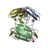
| ||||||||
|---|---|---|---|---|---|---|---|---|---|
| 1 |
| ||||||||
| Unit cell |
|
-Components
| #1: Protein | Mass: 11734.707 Da / Num. of mol.: 4 Source method: isolated from a genetically manipulated source Source: (gene. exp.) PSEUDOMONAS AERUGINOSA (bacteria) / Strain: PAO1 / Plasmid: PET25PAIIL / Production host: #2: Sugar | ChemComp-GXL /   Type: L-saccharide, alpha linking / Mass: 180.156 Da / Num. of mol.: 4 Type: L-saccharide, alpha linking / Mass: 180.156 Da / Num. of mol.: 4Source method: isolated from a genetically manipulated source Formula: C6H12O6 #3: Chemical | ChemComp-CA /   Mass: 40.078 Da / Num. of mol.: 8 / Source method: obtained synthetically / Formula: Ca Mass: 40.078 Da / Num. of mol.: 8 / Source method: obtained synthetically / Formula: Ca#4: Chemical |   Mass: 96.063 Da / Num. of mol.: 2 / Source method: obtained synthetically / Formula: SO4 Mass: 96.063 Da / Num. of mol.: 2 / Source method: obtained synthetically / Formula: SO4#5: Water | ChemComp-HOH / |  Mass: 18.015 Da / Num. of mol.: 430 / Source method: isolated from a natural source / Formula: H2O Mass: 18.015 Da / Num. of mol.: 430 / Source method: isolated from a natural source / Formula: H2O |
|---|
-Experimental details
-Experiment
| Experiment | Method: X-RAY DIFFRACTION / Number of used crystals: 1 |
|---|
- Sample preparation
| Crystal | Density Matthews: 1.9 Å3/Da / Density % sol: 34.72 % |
|---|---|
| Crystal grow | pH: 8.5 Details: TRIS HCL 0.1M, PH8.5, 1.75 M AMMONIUM SULFATE, pH 8.50 |
-Data collection
| Diffraction | Mean temperature: 100 K |
|---|---|
| Diffraction source | Source: SYNCHROTRON / Site: ESRF  / Beamline: ID14-4 / Wavelength: 0.932 / Beamline: ID14-4 / Wavelength: 0.932 |
| Detector | Type: ADSC CCD / Detector: CCD / Date: Mar 17, 2003 / Details: MIRROR |
| Radiation | Monochromator: SINGLE CRYSTAL DIAMOND / Protocol: SINGLE WAVELENGTH / Monochromatic (M) / Laue (L): M / Scattering type: x-ray |
| Radiation wavelength | Wavelength: 0.932 Å / Relative weight: 1 |
| Reflection | Resolution: 1.5→19.43 Å / Num. obs: 63528 / % possible obs: 97 % / Redundancy: 3.82 % / Biso Wilson estimate: 10.28 Å2 / Rmerge(I) obs: 0.05 / Net I/σ(I): 10.35 |
| Reflection shell | Resolution: 1.5→1.55 Å / Redundancy: 3.8 % / Rmerge(I) obs: 0.23 / Mean I/σ(I) obs: 3.08 / % possible all: 95.1 |
- Processing
| Software |
| ||||||||||||||||||||||||||||||||||||||||||||||||||||||||||||||||||||||||||||||||||||||||||||||||||||||||||||||||||||||||||||||||||||||||||||||||||||||||||||||||||||||||||||||||||||||
|---|---|---|---|---|---|---|---|---|---|---|---|---|---|---|---|---|---|---|---|---|---|---|---|---|---|---|---|---|---|---|---|---|---|---|---|---|---|---|---|---|---|---|---|---|---|---|---|---|---|---|---|---|---|---|---|---|---|---|---|---|---|---|---|---|---|---|---|---|---|---|---|---|---|---|---|---|---|---|---|---|---|---|---|---|---|---|---|---|---|---|---|---|---|---|---|---|---|---|---|---|---|---|---|---|---|---|---|---|---|---|---|---|---|---|---|---|---|---|---|---|---|---|---|---|---|---|---|---|---|---|---|---|---|---|---|---|---|---|---|---|---|---|---|---|---|---|---|---|---|---|---|---|---|---|---|---|---|---|---|---|---|---|---|---|---|---|---|---|---|---|---|---|---|---|---|---|---|---|---|---|---|---|---|
| Refinement | Method to determine structure: MOLECULAR REPLACEMENT Starting model: PDB ENTRY 1UZV Resolution: 1.5→19.43 Å / Cor.coef. Fo:Fc: 0.969 / Cor.coef. Fo:Fc free: 0.961 / SU B: 0.992 / SU ML: 0.038 / Cross valid method: THROUGHOUT / ESU R: 0.064 / ESU R Free: 0.064 / Stereochemistry target values: MAXIMUM LIKELIHOOD / Details: HYDROGENS HAVE BEEN ADDED IN THE RIDING POSITIONS.
| ||||||||||||||||||||||||||||||||||||||||||||||||||||||||||||||||||||||||||||||||||||||||||||||||||||||||||||||||||||||||||||||||||||||||||||||||||||||||||||||||||||||||||||||||||||||
| Solvent computation | Ion probe radii: 0.8 Å / Shrinkage radii: 0.8 Å / VDW probe radii: 1.2 Å / Solvent model: MASK | ||||||||||||||||||||||||||||||||||||||||||||||||||||||||||||||||||||||||||||||||||||||||||||||||||||||||||||||||||||||||||||||||||||||||||||||||||||||||||||||||||||||||||||||||||||||
| Displacement parameters | Biso mean: 10.22 Å2
| ||||||||||||||||||||||||||||||||||||||||||||||||||||||||||||||||||||||||||||||||||||||||||||||||||||||||||||||||||||||||||||||||||||||||||||||||||||||||||||||||||||||||||||||||||||||
| Refinement step | Cycle: LAST / Resolution: 1.5→19.43 Å
| ||||||||||||||||||||||||||||||||||||||||||||||||||||||||||||||||||||||||||||||||||||||||||||||||||||||||||||||||||||||||||||||||||||||||||||||||||||||||||||||||||||||||||||||||||||||
| Refine LS restraints |
|