 Movie
Movie Controller

[English] 日本語
 Yorodumi
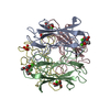
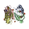
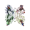
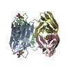
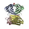
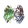
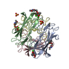
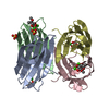
- PDB-1uzv: High affinity fucose binding of Pseudomonas aeruginosa lectin II:... -
+ Open data

- Basic information
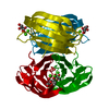
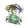
| Entry | Database: PDB / ID: 1uzv | ||||||
|---|---|---|---|---|---|---|---|
| Title | High affinity fucose binding of Pseudomonas aeruginosa lectin II: 1.0 A crystal structure of the complex | ||||||
 Components Components | PSEUDOMONAS AERUGINOSA LECTIN II | ||||||
 Keywords Keywords | LECTIN / FUCOSE / CALCIUM | ||||||
| Function / homology |  Function and homology information Function and homology informationsingle-species biofilm formation / carbohydrate binding / metal ion binding Similarity search - Function | ||||||
| Biological species |   PSEUDOMONAS AERUGINOSA (bacteria) PSEUDOMONAS AERUGINOSA (bacteria) | ||||||
| Method |  X-RAY DIFFRACTION / SYNCHROTRON / MOLECULAR REPLACEMENT / Resolution: 1 Å X-RAY DIFFRACTION / SYNCHROTRON / MOLECULAR REPLACEMENT / Resolution: 1 Å | ||||||
 Authors Authors | Mitchell, E. / Sabin, C.D. / Snajdrova, L. / Budova, M. / Perret, S. / Gautier, C. / Gilboa-Garber, N. / Koca, J. / Wimmerova, M. / Imberty, A. | ||||||
 Citation Citation | Journal: Proteins / Year: 2005 Title: High Affinity Fucose Binding of Pseudomonas Aeruginosa Lectin Pa-Iil: 1.0 A Resolution Crystal Structure of the Complex Combined with Thermodynamics and Computational Chemistry Approaches. Authors: Mitchell, E.P. / Sabin, C. / Snajdrova, L. / Pokorna, M. / Perret, S. / Gautier, C. / Hofr, C. / Gilboa-Garber, N. / Koca, J. / Wimmerova, M. / Imberty, A. #1: Journal: Nat.Struct.Biol. / Year: 2002Title: Structural Basis for Oligosaccharide-Mediated Adhesion of Pseudomonas Aeruginosa in the Lungs of Cystic Fibrosis Patients Authors: Mitchell, E. / Houles, C. / Sudakevitz, D. / Wimmerova, M. / Gautier, C. / Perez, S. / Wu, A.M. / Gilboa-Garber, N. / Imberty, A. | ||||||
| History |
| ||||||
| Remark 700 | SHEET DETERMINATION METHOD: AUTHOR PROVIDED. |
- Structure visualization
| Structure viewer | Molecule: MolmilJmol/JSmol |
|---|
- Downloads & links
-Download
| PDBx/mmCIF format | 1uzv.cif.gz | 199.9 KB | Display | PDBx/mmCIF format |
|---|---|---|---|---|
| PDB format | pdb1uzv.ent.gz | 159.9 KB | Display | PDB format |
| PDBx/mmJSON format | 1uzv.json.gz | Tree view | PDBx/mmJSON format | |
| Others |  Other downloads Other downloads |
-Validation report
| Arichive directory | https://data.pdbj.org/pub/pdb/validation_reports/uz/1uzvftp://data.pdbj.org/pub/pdb/validation_reports/uz/1uzv | HTTPS FTP |
|---|
-Related structure data


-Links
 PDBj
PDBj
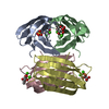
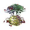
- Assembly
| Deposited unit | 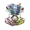
| ||||||||
|---|---|---|---|---|---|---|---|---|---|
| 1 |
| ||||||||
| Unit cell |
|
-Components
| #1: Protein | Mass: 11734.707 Da / Num. of mol.: 4 / Source method: isolated from a natural source / Source: (natural) PSEUDOMONAS AERUGINOSA (bacteria) / References: UniProt: Q9HYN5#2: Chemical |   Mass: 96.063 Da / Num. of mol.: 2 / Source method: obtained synthetically / Formula: SO4 Mass: 96.063 Da / Num. of mol.: 2 / Source method: obtained synthetically / Formula: SO4#3: Chemical | ChemComp-CA /   Mass: 40.078 Da / Num. of mol.: 8 / Source method: obtained synthetically / Formula: Ca Mass: 40.078 Da / Num. of mol.: 8 / Source method: obtained synthetically / Formula: Ca#4: Sugar | ChemComp-FUC /   Type: L-saccharide, alpha linking / Mass: 164.156 Da / Num. of mol.: 4 Type: L-saccharide, alpha linking / Mass: 164.156 Da / Num. of mol.: 4Source method: isolated from a genetically manipulated source Formula: C6H12O5 #5: Water | ChemComp-HOH / |  Mass: 18.015 Da / Num. of mol.: 682 / Source method: isolated from a natural source / Formula: H2O Mass: 18.015 Da / Num. of mol.: 682 / Source method: isolated from a natural source / Formula: H2O |
|---|
-Experimental details
-Experiment
| Experiment | Method: X-RAY DIFFRACTION / Number of used crystals: 1 |
|---|
- Sample preparation
| Crystal | Density Matthews: 1.87 Å3/Da / Density % sol: 31.1 % Description: MOLECULAR REPLACEMENT WAS USED TO DETERMINE CA ION POSITIONS AND SUBSEQUENT AB INITIO PHASING. DATA WERE COLLECTED IN TWO SECTIONS, ONE SWEEP FOR HIGH RESOLUTION DATA AND ANOTHER FOR LOW RESOLUTION DATA. |
|---|---|
| Crystal grow | pH: 8.5 Details: TRIS HCL 0.1M, PH8.5, 1.75 M AMMONIUM SULFATE, pH 8.50 |
-Data collection
| Diffraction | Mean temperature: 100 K |
|---|---|
| Diffraction source | Source: SYNCHROTRON / Site: ESRF  / Beamline: ID14-2 / Wavelength: 0.9326 / Beamline: ID14-2 / Wavelength: 0.9326 |
| Detector | Type: ADSC CCD / Detector: CCD / Date: Jan 15, 2002 / Details: TOROIDAL MIRROR |
| Radiation | Monochromator: SINGLE CRYSTAL DIAMOND / Protocol: SINGLE WAVELENGTH / Monochromatic (M) / Laue (L): M / Scattering type: x-ray |
| Radiation wavelength | Wavelength: 0.9326 Å / Relative weight: 1 |
| Reflection | Resolution: 1→29.88 Å / Num. obs: 216831 / % possible obs: 97.7 % / Redundancy: 3.3 % / Rmerge(I) obs: 0.063 / Net I/σ(I): 5 |
| Reflection shell | Resolution: 1→1.04 Å / Redundancy: 2.9 % / Rmerge(I) obs: 0.23 / Mean I/σ(I) obs: 2.8 / % possible all: 94.3 |
- Processing
| Software |
| |||||||||||||||||||||||||||||||||
|---|---|---|---|---|---|---|---|---|---|---|---|---|---|---|---|---|---|---|---|---|---|---|---|---|---|---|---|---|---|---|---|---|---|---|
| Refinement | Method to determine structure: MOLECULAR REPLACEMENT Starting model: PSEUDOMONAS AERUGINOSA LECTIN Resolution: 1→30 Å / Num. parameters: 35194 / Num. restraintsaints: 45801 / Cross valid method: FREE R-VALUE / σ(F): 0 / Stereochemistry target values: ENGH AND HUBER Details: INITIAL REFINEMENT WITH REFMAC5 AND THEN SHELX-97 WITH ANISOTROPIC REFINEMENT USING ALL REFLECTIONS (NO CUTOFF)
| |||||||||||||||||||||||||||||||||
| Solvent computation | Solvent model: MOEWS & KRETSINGER, J.MOL.BIOL.91(1973)201-2 | |||||||||||||||||||||||||||||||||
| Refine analyze | Num. disordered residues: 27 / Occupancy sum hydrogen: 2991.64 / Occupancy sum non hydrogen: 4024.72 | |||||||||||||||||||||||||||||||||
| Refinement step | Cycle: LAST / Resolution: 1→30 Å
| |||||||||||||||||||||||||||||||||
| Refine LS restraints |
|
