- PDB-4c93: Crystal structure of the carboxy-terminal domain of yeast Ctf4 bo... -
+
データを開く
IDまたはキーワード:
読み込み中...
-
基本情報
登録情報
データベース: PDB / ID: 4c93
タイトル
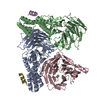
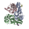
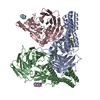
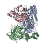
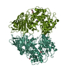
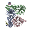
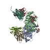
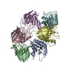
Crystal structure of the carboxy-terminal domain of yeast Ctf4 bound to Pol alpha.
要素
DNA POLYMERASE ALPHA CATALYTIC SUBUNIT A
DNA POLYMERASE ALPHA-BINDING PROTEIN
キーワード
DNA REPLICATION / ADAPTOR PROTEIN / BETA PROPELLER DOMAIN
機能・相同性
機能・相同性情報
Inhibition of replication initiation of damaged DNA by RB1/E2F1 / establishment of sister chromatid cohesion / H3-H4 histone complex chaperone activity / DNA replication initiation / Processive synthesis on the lagging strand / RNA-templated DNA biosynthetic process / Removal of the Flap Intermediate / Polymerase switching / premeiotic DNA replication / alpha DNA polymerase:primase complex ...Inhibition of replication initiation of damaged DNA by RB1/E2F1 / establishment of sister chromatid cohesion / H3-H4 histone complex chaperone activity / DNA replication initiation / Processive synthesis on the lagging strand / RNA-templated DNA biosynthetic process / Removal of the Flap Intermediate / Polymerase switching / premeiotic DNA replication / alpha DNA polymerase:primase complex / Activation of the pre-replicative complex / lagging strand elongation / double-strand break repair via break-induced replication / mitotic DNA replication initiation / mitotic sister chromatid cohesion / DNA synthesis involved in DNA repair / leading strand elongation / nuclear chromosome / nuclear replication fork / DNA replication origin binding / DNA replication initiation / replication fork / DNA-templated DNA replication / double-strand break repair / mitotic cell cycle / nucleosome assembly / single-stranded DNA binding / 4 iron, 4 sulfur cluster binding / DNA-directed DNA polymerase / DNA-directed DNA polymerase activity / DNA replication / nucleotide binding / DNA repair / chromatin binding / mitochondrion / zinc ion binding / identical protein binding / nucleus 類似検索 - 分子機能
: / DNA polymerase alpha-binding protein Ctf4, C-terminal domain / Minichromosome loss protein Mcl1, middle region / Minichromosome loss protein, Mcl1, middle region / DNA polymerase alpha catalytic subunit, N-terminal domain / DNA polymerase alpha, zinc finger domain superfamily / DNA Polymerase alpha zinc finger / DNA polymerase alpha subunit p180 N terminal / Zinc finger, DNA-directed DNA polymerase, family B, alpha / DNA polymerase alpha catalytic subunit, catalytic domain ...: / DNA polymerase alpha-binding protein Ctf4, C-terminal domain / Minichromosome loss protein Mcl1, middle region / Minichromosome loss protein, Mcl1, middle region / DNA polymerase alpha catalytic subunit, N-terminal domain / DNA polymerase alpha, zinc finger domain superfamily / DNA Polymerase alpha zinc finger / DNA polymerase alpha subunit p180 N terminal / Zinc finger, DNA-directed DNA polymerase, family B, alpha / DNA polymerase alpha catalytic subunit, catalytic domain / DNA polymerase family B, thumb domain / DNA-directed DNA polymerase, family B, multifunctional domain / DNA-directed DNA polymerase, family B, conserved site / DNA polymerase family B signature. / DNA polymerase family B / DNA polymerase family B, exonuclease domain / DNA-directed DNA polymerase, family B, exonuclease domain / DNA polymerase, palm domain superfamily / DNA polymerase type-B family / DNA-directed DNA polymerase, family B / Ribonuclease H superfamily / Ribonuclease H-like superfamily / WD40-repeat-containing domain superfamily / WD40/YVTN repeat-like-containing domain superfamily / DNA/RNA polymerase superfamily 類似検索 - ドメイン・相同性
DNA polymerase alpha catalytic subunit A / DNA polymerase alpha-binding protein 類似検索 - 構成要素
#241 - 2020年1月 20年の分子を振り返って (Twenty Years of Molecules) 類似性 (3)
-
集合体
登録構造単位
A: DNA POLYMERASE ALPHA-BINDING PROTEIN B: DNA POLYMERASE ALPHA-BINDING PROTEIN C: DNA POLYMERASE ALPHA-BINDING PROTEIN D: DNA POLYMERASE ALPHA CATALYTIC SUBUNIT A E: DNA POLYMERASE ALPHA CATALYTIC SUBUNIT A
DNAPOLYMERASEALPHACATALYTICSUBUNITA / DNA POLYMERASE I SUBUNIT A / DNA POLYMERASE ALPHA\: PRIMASE COMPLEX P180 SUBUNIT / DNA POLYMERASE- ...DNA POLYMERASE I SUBUNIT A / DNA POLYMERASE ALPHA\: PRIMASE COMPLEX P180 SUBUNIT / DNA POLYMERASE-PRIMASE COMPLEX P180 SUBUNIT / POL ALPHA-PRIMASE COMPLEX P180 SUBUNIT / DNA POLYMERASE ALPHA CATALYTIC SUBUNIT
 ムービー
ムービー コントローラー


 データを開く
 基本情報
基本情報 要素
要素 キーワード
キーワード 機能・相同性情報
 X線回折 /
  データ登録者
 引用
引用 構造の表示
構造の表示 ダウンロードとリンク
 その他のダウンロード


 PDBj
PDBj


 集合体
集合体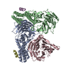

 分子量: 18.015 Da / 分子数: 160 / 由来タイプ: 天然 / 式: H2O
 試料調製
試料調製 / ビームライン: I04 / 波長: 0.9795
  解析
解析