 Movie
Movie Controller

[English] 日本語
 Yorodumi
- PDB-5hoi: Crystal structure of the carboxy-terminal domain of yeast Ctf4 bo... -
+ Open data

- Basic information
| Entry | Database: PDB / ID: 5hoi | |||||||||
|---|---|---|---|---|---|---|---|---|---|---|
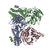
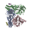
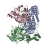
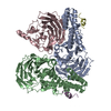
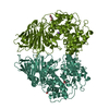
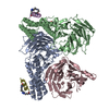
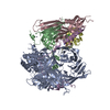
| Title | Crystal structure of the carboxy-terminal domain of yeast Ctf4 bound to Tof2. | |||||||||
 Components Components |
| |||||||||
 Keywords Keywords | REPLICATION / DNA replication / adaptor protein / beta-propeller domain | |||||||||
| Function / homology |  Function and homology information Function and homology informationestablishment of sister chromatid cohesion / protein localization to nucleolar rDNA repeats / rDNA chromatin condensation / phosphatase activator activity / rDNA binding / rDNA heterochromatin formation / double-strand break repair via break-induced replication / mitotic sister chromatid cohesion / nucleolus organization / nuclear chromosome ...establishment of sister chromatid cohesion / protein localization to nucleolar rDNA repeats / rDNA chromatin condensation / phosphatase activator activity / rDNA binding / rDNA heterochromatin formation / double-strand break repair via break-induced replication / mitotic sister chromatid cohesion / nucleolus organization / nuclear chromosome / nuclear replication fork / DNA replication initiation / DNA-templated DNA replication / mitotic cell cycle / nucleosome assembly / DNA repair / chromatin binding / nucleolus / mitochondrion / identical protein binding / nucleus Similarity search - Function | |||||||||
| Biological species |  | |||||||||
| Method |  X-RAY DIFFRACTION / SYNCHROTRON / Resolution: 3.3 Å X-RAY DIFFRACTION / SYNCHROTRON / Resolution: 3.3 Å | |||||||||
 Authors Authors | Simon, A.C. / Pellegrini, L. | |||||||||
| Funding support |  United Kingdom, 2items United Kingdom, 2items
| |||||||||
 Citation Citation | Journal: Mol.Cell / Year: 2016 Title: Ctf4 Is a Hub in the Eukaryotic Replisome that Links Multiple CIP-Box Proteins to the CMG Helicase. Authors: Villa, F. / Simon, A.C. / Ortiz Bazan, M.A. / Kilkenny, M.L. / Wirthensohn, D. / Wightman, M. / Matak-Vinkovic, D. / Pellegrini, L. / Labib, K. #1: Journal: Nature / Year: 2014Title: A Ctf4 trimer couples the CMG helicase to DNA polymerase alpha in the eukaryotic replisome. Authors: Simon, A.C. / Zhou, J.C. / Perera, R.L. / van Deursen, F. / Evrin, C. / Ivanova, M.E. / Kilkenny, M.L. / Renault, L. / Kjaer, S. / Matak-Vinkovic, D. / Labib, K. / Costa, A. / Pellegrini, L. | |||||||||
| History |
|
- Structure visualization
| Structure viewer | Molecule: MolmilJmol/JSmol |
|---|
- Downloads & links
-Download
| PDBx/mmCIF format | 5hoi.cif.gz | 251.2 KB | Display | PDBx/mmCIF format |
|---|---|---|---|---|
| PDB format | pdb5hoi.ent.gz | 200.4 KB | Display | PDB format |
| PDBx/mmJSON format | 5hoi.json.gz | Tree view | PDBx/mmJSON format | |
| Others |  Other downloads Other downloads |
-Validation report
| Arichive directory | https://data.pdbj.org/pub/pdb/validation_reports/ho/5hoiftp://data.pdbj.org/pub/pdb/validation_reports/ho/5hoi | HTTPS FTP |
|---|
-Related structure data


-Links
 PDBj
PDBj- Assembly
| Deposited unit | 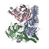
| ||||||||
|---|---|---|---|---|---|---|---|---|---|
| 1 |
| ||||||||
| Unit cell |
|
-Components
| #1: Protein | Mass: 54447.484 Da / Num. of mol.: 3 / Fragment: UNP residues 472-927 Source method: isolated from a genetically manipulated source Source: (gene. exp.) Gene: CTF4, CHL15, POB1, YPR135W, P9659.7 / Production host:  #2: Protein/peptide | Mass: 2516.958 Da / Num. of mol.: 3 / Fragment: UNP residues 497-517 / Source method: obtained synthetically Details: The synthetic peptide corresponds to amino acids 497 to 517 of yeast Tof2. Source: (synth.) #3: Water | ChemComp-HOH / |  Mass: 18.015 Da / Num. of mol.: 51 / Source method: isolated from a natural source / Formula: H2O Mass: 18.015 Da / Num. of mol.: 51 / Source method: isolated from a natural source / Formula: H2O |
|---|
-Experimental details
-Experiment
| Experiment | Method: X-RAY DIFFRACTION |
|---|
- Sample preparation
| Crystal | Density Matthews: 2.82 Å3/Da / Density % sol: 56.39 % |
|---|---|
| Crystal grow | Temperature: 292 K / Method: vapor diffusion, hanging drop Details: 0.2 M tri-sodium citrate pH 6.2, 7-9% PEG 8000 and 0.45-0.9 M NaCl |
-Data collection
| Diffraction | Mean temperature: 100 K |
|---|---|
| Diffraction source | Source: SYNCHROTRON / Site: Diamond / Beamline: I02 / Wavelength: 0.97949 Å |
| Detector | Type: PSI PILATUS 6M / Detector: PIXEL / Date: May 13, 2015 |
| Radiation | Protocol: SINGLE WAVELENGTH / Monochromatic (M) / Laue (L): M / Scattering type: x-ray |
| Radiation wavelength | Wavelength: 0.97949 Å / Relative weight: 1 |
| Reflection | Resolution: 3.3→48.99 Å / Num. obs: 29749 / % possible obs: 99.3 % / Redundancy: 4.6 % / Net I/σ(I): 8.2 |
| Reflection shell | Resolution: 3.3→3.5 Å / Redundancy: 4.5 % / Mean I/σ(I) obs: 2.1 / % possible all: 98.5 |
- Processing
| Software |
| |||||||||||||||||||||||||||||||||||||||||||||||||||||||||||||||||||||||||||||||||||||||||||||||||||||||||||||||||||||||||||||||||||||||||||||||||||
|---|---|---|---|---|---|---|---|---|---|---|---|---|---|---|---|---|---|---|---|---|---|---|---|---|---|---|---|---|---|---|---|---|---|---|---|---|---|---|---|---|---|---|---|---|---|---|---|---|---|---|---|---|---|---|---|---|---|---|---|---|---|---|---|---|---|---|---|---|---|---|---|---|---|---|---|---|---|---|---|---|---|---|---|---|---|---|---|---|---|---|---|---|---|---|---|---|---|---|---|---|---|---|---|---|---|---|---|---|---|---|---|---|---|---|---|---|---|---|---|---|---|---|---|---|---|---|---|---|---|---|---|---|---|---|---|---|---|---|---|---|---|---|---|---|---|---|---|---|
| Refinement | Resolution: 3.3→48.99 Å / SU ML: 0.41 / Cross valid method: FREE R-VALUE / σ(F): 0.08 / Phase error: 22.64 / Stereochemistry target values: ML
| |||||||||||||||||||||||||||||||||||||||||||||||||||||||||||||||||||||||||||||||||||||||||||||||||||||||||||||||||||||||||||||||||||||||||||||||||||
| Solvent computation | Shrinkage radii: 0.9 Å / VDW probe radii: 1.11 Å / Solvent model: FLAT BULK SOLVENT MODEL | |||||||||||||||||||||||||||||||||||||||||||||||||||||||||||||||||||||||||||||||||||||||||||||||||||||||||||||||||||||||||||||||||||||||||||||||||||
| Refinement step | Cycle: LAST / Resolution: 3.3→48.99 Å
| |||||||||||||||||||||||||||||||||||||||||||||||||||||||||||||||||||||||||||||||||||||||||||||||||||||||||||||||||||||||||||||||||||||||||||||||||||
| Refine LS restraints |
| |||||||||||||||||||||||||||||||||||||||||||||||||||||||||||||||||||||||||||||||||||||||||||||||||||||||||||||||||||||||||||||||||||||||||||||||||||
| LS refinement shell |
|
