 Movie
Movie Controller

[English] 日本語
 Yorodumi
- PDB-4bzz: Complete crystal structure of carboxylesterase Cest-2923 from Lac... -
+ Open data

- Basic information
| Entry | Database: PDB / ID: 4bzz | ||||||
|---|---|---|---|---|---|---|---|
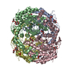
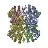
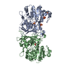
| Title | Complete crystal structure of carboxylesterase Cest-2923 from Lactobacillus plantarum WCFS1 | ||||||
 Components Components | LIPASE/ESTERASE | ||||||
 Keywords Keywords | HYDROLASE / CARBOXYLESTERASE | ||||||
| Function / homology |  Function and homology information Function and homology information | ||||||
| Biological species |  LACTOBACILLUS PLANTARUM (bacteria) LACTOBACILLUS PLANTARUM (bacteria) | ||||||
| Method |  X-RAY DIFFRACTION / SYNCHROTRON / MOLECULAR REPLACEMENT / Resolution: 3 Å X-RAY DIFFRACTION / SYNCHROTRON / MOLECULAR REPLACEMENT / Resolution: 3 Å | ||||||
 Authors Authors | Benavente, R. / Esteban-Torres, M. / Acebron, I. / de las Rivas, B. / Munoz, R. / Alvarez, Y. / Mancheno, J.M. | ||||||
 Citation Citation | Journal: FEBS J. / Year: 2013 Title: Structure, Biochemical Characterization and Analysis of the Pleomorphism of Carboxylesterase Cest-2923 from Lactobacillus Plantarum Wcfs1 Authors: Benavente, R. / Esteban-Torres, M. / Acebron, I. / De Las Rivas, B. / Munoz, R. / Alvarez, Y. / Mancheno, J.M. | ||||||
| History |
|
- Structure visualization
| Structure viewer | Molecule: MolmilJmol/JSmol |
|---|
- Downloads & links
-Download
| PDBx/mmCIF format | 4bzz.cif.gz | 122 KB | Display | PDBx/mmCIF format |
|---|---|---|---|---|
| PDB format | pdb4bzz.ent.gz | 95.9 KB | Display | PDB format |
| PDBx/mmJSON format | 4bzz.json.gz | Tree view | PDBx/mmJSON format | |
| Others |  Other downloads Other downloads |
-Validation report
| Arichive directory | https://data.pdbj.org/pub/pdb/validation_reports/bz/4bzzftp://data.pdbj.org/pub/pdb/validation_reports/bz/4bzz | HTTPS FTP |
|---|
-Related structure data
| Related structure data | 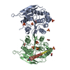 4bzwC 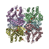 4c01C 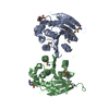 3d3nS C: citing same article ( S: Starting model for refinement |
|---|---|
| Similar structure data |


-Links
 PDBj
PDBj- Assembly
| Deposited unit | 
| ||||||||
|---|---|---|---|---|---|---|---|---|---|
| 1 | x 6
| ||||||||
| Unit cell |
|
-Components
| #1: Protein | Mass: 31275.307 Da / Num. of mol.: 1 Source method: isolated from a genetically manipulated source Source: (gene. exp.) LACTOBACILLUS PLANTARUM (bacteria) / Strain: WCFS1 / Production host: | ||||||
|---|---|---|---|---|---|---|---|
| #2: Chemical |   Mass: 59.044 Da / Num. of mol.: 3 / Source method: obtained synthetically / Formula: C2H3O2 Mass: 59.044 Da / Num. of mol.: 3 / Source method: obtained synthetically / Formula: C2H3O2#3: Chemical | ChemComp-SO4 /   Mass: 96.063 Da / Num. of mol.: 4 / Source method: obtained synthetically / Formula: SO4 Mass: 96.063 Da / Num. of mol.: 4 / Source method: obtained synthetically / Formula: SO4#4: Chemical | ChemComp-CCN / |   Mass: 41.052 Da / Num. of mol.: 1 / Source method: obtained synthetically / Formula: C2H3N Mass: 41.052 Da / Num. of mol.: 1 / Source method: obtained synthetically / Formula: C2H3N#5: Water | ChemComp-HOH / |  Mass: 18.015 Da / Num. of mol.: 4 / Source method: isolated from a natural source / Formula: H2O Mass: 18.015 Da / Num. of mol.: 4 / Source method: isolated from a natural source / Formula: H2O |
-Experimental details
-Experiment
| Experiment | Method: X-RAY DIFFRACTION / Number of used crystals: 1 |
|---|
- Sample preparation
| Crystal | Density Matthews: 3.8 Å3/Da / Density % sol: 67 % / Description: NONE |
|---|---|
| Crystal grow | pH: 4.6 Details: 1.7 M AMMONIUM SULPHATE, 0.15 M SODIUM ACETATE, PH 4.6 |
-Data collection
| Diffraction | Mean temperature: 100 K |
|---|---|
| Diffraction source | Source: SYNCHROTRON / Site: ESRF  / Beamline: ID23-1 / Wavelength: 0.9687 / Beamline: ID23-1 / Wavelength: 0.9687 |
| Detector | Type: ADSC CCD / Detector: CCD |
| Radiation | Protocol: SINGLE WAVELENGTH / Monochromatic (M) / Laue (L): M / Scattering type: x-ray |
| Radiation wavelength | Wavelength: 0.9687 Å / Relative weight: 1 |
| Reflection | Resolution: 2.99→49 Å / Num. obs: 9994 / % possible obs: 99.6 % / Redundancy: 7.5 % / Biso Wilson estimate: 52.06 Å2 / Rmerge(I) obs: 0.12 / Net I/σ(I): 13.7 |
| Reflection shell | Resolution: 2.99→3.15 Å / Redundancy: 7.3 % / Rmerge(I) obs: 0.47 / Mean I/σ(I) obs: 4.1 / % possible all: 98.9 |
- Processing
| Software |
| |||||||||||||||||||||||||||||||||||||||||||||||||||||||||||||||||||||||||||||||||||||||||||||||||||||||||||||||||||||||||||||
|---|---|---|---|---|---|---|---|---|---|---|---|---|---|---|---|---|---|---|---|---|---|---|---|---|---|---|---|---|---|---|---|---|---|---|---|---|---|---|---|---|---|---|---|---|---|---|---|---|---|---|---|---|---|---|---|---|---|---|---|---|---|---|---|---|---|---|---|---|---|---|---|---|---|---|---|---|---|---|---|---|---|---|---|---|---|---|---|---|---|---|---|---|---|---|---|---|---|---|---|---|---|---|---|---|---|---|---|---|---|---|---|---|---|---|---|---|---|---|---|---|---|---|---|---|---|---|
| Refinement | Method to determine structure: MOLECULAR REPLACEMENT Starting model: PDB EBTRY 3D3N Resolution: 3→46.062 Å / SU ML: 0.26 / σ(F): 1.34 / Phase error: 19.67 / Stereochemistry target values: ML
| |||||||||||||||||||||||||||||||||||||||||||||||||||||||||||||||||||||||||||||||||||||||||||||||||||||||||||||||||||||||||||||
| Solvent computation | Shrinkage radii: 0.9 Å / VDW probe radii: 1.11 Å / Solvent model: FLAT BULK SOLVENT MODEL | |||||||||||||||||||||||||||||||||||||||||||||||||||||||||||||||||||||||||||||||||||||||||||||||||||||||||||||||||||||||||||||
| Refinement step | Cycle: LAST / Resolution: 3→46.062 Å
| |||||||||||||||||||||||||||||||||||||||||||||||||||||||||||||||||||||||||||||||||||||||||||||||||||||||||||||||||||||||||||||
| Refine LS restraints |
| |||||||||||||||||||||||||||||||||||||||||||||||||||||||||||||||||||||||||||||||||||||||||||||||||||||||||||||||||||||||||||||
| LS refinement shell |
| |||||||||||||||||||||||||||||||||||||||||||||||||||||||||||||||||||||||||||||||||||||||||||||||||||||||||||||||||||||||||||||
| Refinement TLS params. | Method: refined / Refine-ID: X-RAY DIFFRACTION
| |||||||||||||||||||||||||||||||||||||||||||||||||||||||||||||||||||||||||||||||||||||||||||||||||||||||||||||||||||||||||||||
| Refinement TLS group |
|
