 Movie
Movie Controller

[English] 日本語
 Yorodumi
- PDB-4bsz: Crystal Structure of the Yeast Ribosomal Protein Rps3 in Complex ... -
+ Open data

- Basic information
| Entry | Database: PDB / ID: 4bsz | ||||||
|---|---|---|---|---|---|---|---|
| Title | Crystal Structure of the Yeast Ribosomal Protein Rps3 in Complex with its Chaperone Yar1 | ||||||
 Components Components |
| ||||||
 Keywords Keywords | RNA BINDING PROTEIN | ||||||
| Function / homology |  Function and homology information Function and homology informationMBF transcription complex / SBF transcription complex / response to osmotic stress / PELO:HBS1L and ABCE1 dissociate a ribosome on a non-stop mRNA / nonfunctional rRNA decay / Formation of the ternary complex, and subsequently, the 43S complex / Translation initiation complex formation / Ribosomal scanning and start codon recognition / preribosome, small subunit precursor / Major pathway of rRNA processing in the nucleolus and cytosol ...MBF transcription complex / SBF transcription complex / response to osmotic stress / PELO:HBS1L and ABCE1 dissociate a ribosome on a non-stop mRNA / nonfunctional rRNA decay / Formation of the ternary complex, and subsequently, the 43S complex / Translation initiation complex formation / Ribosomal scanning and start codon recognition / preribosome, small subunit precursor / Major pathway of rRNA processing in the nucleolus and cytosol / SRP-dependent cotranslational protein targeting to membrane / GTP hydrolysis and joining of the 60S ribosomal subunit / Formation of a pool of free 40S subunits / Nonsense Mediated Decay (NMD) independent of the Exon Junction Complex (EJC) / Nonsense Mediated Decay (NMD) enhanced by the Exon Junction Complex (EJC) / L13a-mediated translational silencing of Ceruloplasmin expression / 90S preribosome / ribosomal subunit export from nucleus / ribosomal small subunit export from nucleus / DNA-(apurinic or apyrimidinic site) endonuclease activity / maintenance of translational fidelity / : / regulation of protein localization / cellular response to oxidative stress / ribosomal small subunit biogenesis / cytosolic small ribosomal subunit / DNA-binding transcription activator activity, RNA polymerase II-specific / cytoplasmic translation / structural constituent of ribosome / positive regulation of transcription by RNA polymerase II / RNA binding / nucleus / cytosol / cytoplasm Similarity search - Function | ||||||
| Biological species |  | ||||||
| Method |  X-RAY DIFFRACTION / SYNCHROTRON / MOLECULAR REPLACEMENT / Resolution: 2.842 Å X-RAY DIFFRACTION / SYNCHROTRON / MOLECULAR REPLACEMENT / Resolution: 2.842 Å | ||||||
 Authors Authors | Holzer, S. / Ban, N. / Klinge, S. | ||||||
 Citation Citation | Journal: J.Mol.Biol. / Year: 2013 Title: Crystal Structure of the Yeast Ribosomal Protein Rps3 in Complex with its Chaperone Yar1 Authors: Holzer, S. / Ban, N. / Klinge, S. | ||||||
| History |
|
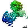
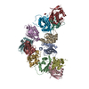
- Structure visualization
| Structure viewer | Molecule: MolmilJmol/JSmol |
|---|
- Downloads & links
-Download
| PDBx/mmCIF format | 4bsz.cif.gz | 147.5 KB | Display | PDBx/mmCIF format |
|---|---|---|---|---|
| PDB format | pdb4bsz.ent.gz | 116.8 KB | Display | PDB format |
| PDBx/mmJSON format | 4bsz.json.gz | Tree view | PDBx/mmJSON format | |
| Others |  Other downloads Other downloads |
-Validation report
| Arichive directory | https://data.pdbj.org/pub/pdb/validation_reports/bs/4bszftp://data.pdbj.org/pub/pdb/validation_reports/bs/4bsz | HTTPS FTP |
|---|
-Related structure data
| Related structure data |  2j00 S: Starting model for refinement |
|---|---|
| Similar structure data |


-Links
 PDBj
PDBj


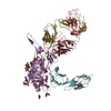
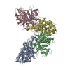
- Assembly
| Deposited unit | 
| ||||||||
|---|---|---|---|---|---|---|---|---|---|
| 1 | 
| ||||||||
| Unit cell |
|
-Components
| #1: Protein | Mass: 26957.266 Da / Num. of mol.: 1 Source method: isolated from a genetically manipulated source Source: (gene. exp.) Strain: W303 / Plasmid: PRSF-DUET1 / Production host:  |
|---|---|
| #2: Protein | Mass: 22608.729 Da / Num. of mol.: 1 Source method: isolated from a genetically manipulated source Source: (gene. exp.) Strain: W303 / Plasmid: PRSF-DUET1 / Production host: |
| Sequence details | THE CRYSTALLISED CONSTRUCT CONTAINS AN N-TERMINAL ADDITION OF THE RESIDUE SEQUENCE MADP AS A RESULT ...THE CRYSTALLIS |
-Experimental details
-Experiment
| Experiment | Method: X-RAY DIFFRACTION / Number of used crystals: 1 |
|---|
- Sample preparation
| Crystal | Density Matthews: 3.53 Å3/Da / Density % sol: 65.19 % Description: SBGRID WIDE SEARCH MOLECULAR REPLACEMENT WSMR, STOKES-REES ET AL 2010, WAS USED AND THE STRUCTURE WAS SOLVED BY USING PHASER WITH THE FOLLOWING ENTRIES 2J00 - CHAIN C, N-TERMINAL DOMAIN 1MJ0A |
|---|---|
| Crystal grow | Details: 0.1 M MES PH 6.0, 200 MM LITHIUM SULPHATE, 20% PEG 4000 |
-Data collection
| Diffraction | Mean temperature: 100 K |
|---|---|
| Diffraction source | Source: SYNCHROTRON / Site: SLS  / Beamline: X06SA / Wavelength: 1 / Beamline: X06SA / Wavelength: 1 |
| Detector | Type: DECTRIS PILATUS 6M / Detector: PIXEL / Date: Dec 12, 2012 |
| Radiation | Protocol: SINGLE WAVELENGTH / Monochromatic (M) / Laue (L): M / Scattering type: x-ray |
| Radiation wavelength | Wavelength: 1 Å / Relative weight: 1 |
| Reflection | Resolution: 2.84→34.57 Å / Num. obs: 12331 / % possible obs: 93.9 % / Observed criterion σ(I): 2 / Redundancy: 8.19130646 % / Biso Wilson estimate: 76.9 Å2 / Rmerge(I) obs: 0.07 / Net I/σ(I): 19.85 |
| Reflection shell | Resolution: 2.84→3.01 Å / Redundancy: 8.41671018 % / Rmerge(I) obs: 0.79 / Mean I/σ(I) obs: 2.86 / % possible all: 94 |
- Processing
| Software |
| ||||||||||||||||||||||||||||||||||||||||||||||||||||||||||||||||||||||||||||||||||||||||||||||||||||||||||||||||||||||||||||||||||||||||||||||||||||||
|---|---|---|---|---|---|---|---|---|---|---|---|---|---|---|---|---|---|---|---|---|---|---|---|---|---|---|---|---|---|---|---|---|---|---|---|---|---|---|---|---|---|---|---|---|---|---|---|---|---|---|---|---|---|---|---|---|---|---|---|---|---|---|---|---|---|---|---|---|---|---|---|---|---|---|---|---|---|---|---|---|---|---|---|---|---|---|---|---|---|---|---|---|---|---|---|---|---|---|---|---|---|---|---|---|---|---|---|---|---|---|---|---|---|---|---|---|---|---|---|---|---|---|---|---|---|---|---|---|---|---|---|---|---|---|---|---|---|---|---|---|---|---|---|---|---|---|---|---|---|---|---|
| Refinement | Method to determine structure: MOLECULAR REPLACEMENT Starting model: PDB ENTRY 2J00 2j00 Resolution: 2.842→34.574 Å / SU ML: 0.4 / σ(F): 2 / Phase error: 23.53 / Stereochemistry target values: ML Details: ABSENT REGIONS INCLUDE THE C-TERMINUS OF RPS3 AND THE C-TERMINUS OF YAR1
| ||||||||||||||||||||||||||||||||||||||||||||||||||||||||||||||||||||||||||||||||||||||||||||||||||||||||||||||||||||||||||||||||||||||||||||||||||||||
| Solvent computation | Shrinkage radii: 0.9 Å / VDW probe radii: 1.11 Å / Solvent model: FLAT BULK SOLVENT MODEL | ||||||||||||||||||||||||||||||||||||||||||||||||||||||||||||||||||||||||||||||||||||||||||||||||||||||||||||||||||||||||||||||||||||||||||||||||||||||
| Displacement parameters | Biso mean: 79.5 Å2 | ||||||||||||||||||||||||||||||||||||||||||||||||||||||||||||||||||||||||||||||||||||||||||||||||||||||||||||||||||||||||||||||||||||||||||||||||||||||
| Refinement step | Cycle: LAST / Resolution: 2.842→34.574 Å
| ||||||||||||||||||||||||||||||||||||||||||||||||||||||||||||||||||||||||||||||||||||||||||||||||||||||||||||||||||||||||||||||||||||||||||||||||||||||
| Refine LS restraints |
| ||||||||||||||||||||||||||||||||||||||||||||||||||||||||||||||||||||||||||||||||||||||||||||||||||||||||||||||||||||||||||||||||||||||||||||||||||||||
| LS refinement shell |
| ||||||||||||||||||||||||||||||||||||||||||||||||||||||||||||||||||||||||||||||||||||||||||||||||||||||||||||||||||||||||||||||||||||||||||||||||||||||
| Refinement TLS params. | Method: refined / Refine-ID: X-RAY DIFFRACTION
| ||||||||||||||||||||||||||||||||||||||||||||||||||||||||||||||||||||||||||||||||||||||||||||||||||||||||||||||||||||||||||||||||||||||||||||||||||||||
| Refinement TLS group |
|
