- PDB-4bf9: Crystal structure of E. coli dihydrouridine synthase C (DusC) (se... -
+
Open data
ID or keywords:
Loading...
-
Basic information
Entry
Database: PDB / ID: 4bf9
Title
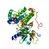
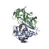
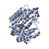
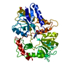
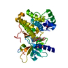
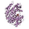
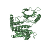
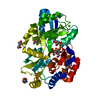
Crystal structure of E. coli dihydrouridine synthase C (DusC) (selenomethionine derivative)
Components
TRNA-DIHYDROURIDINE SYNTHASE C
Keywords
OXIDOREDUCTASE / TRNA MODIFICATION
Function / homology
Function and homology information
tRNA-dihydrouridine16 synthase activity / cellular response to redox state / tRNA dihydrouridine synthesis / tRNA dihydrouridine synthase activity / Oxidoreductases; Acting on the CH-CH group of donors; With NAD+ or NADP+ as acceptor / FMN binding / flavin adenine dinucleotide binding / tRNA binding Similarity search - Function
Dihydrouridine synthase, C-terminal recognition domain / tRNA-dihydrouridine(16) synthase / tRNA-dihydrouridine(16) synthase, C-terminal / tRNA-dihydrouridine synthase / Bacteriocin As-48; Chain A / tRNA-dihydrouridine synthase, conserved site / DUS-like, FMN-binding domain / Dihydrouridine synthase (Dus) / Uncharacterized protein family UPF0034 signature. / Aldolase class I ...Dihydrouridine synthase, C-terminal recognition domain / tRNA-dihydrouridine(16) synthase / tRNA-dihydrouridine(16) synthase, C-terminal / tRNA-dihydrouridine synthase / Bacteriocin As-48; Chain A / tRNA-dihydrouridine synthase, conserved site / DUS-like, FMN-binding domain / Dihydrouridine synthase (Dus) / Uncharacterized protein family UPF0034 signature. / Aldolase class I / Aldolase-type TIM barrel / TIM Barrel / Alpha-Beta Barrel / Up-down Bundle / Mainly Alpha / Alpha Beta Similarity search - Domain/homology
SHEET DETERMINATION METHOD: DSSP THE SHEETS PRESENTED AS "AA" IN EACH CHAIN ON SHEET RECORDS BELOW ... SHEET DETERMINATION METHOD: DSSP THE SHEETS PRESENTED AS "AA" IN EACH CHAIN ON SHEET RECORDS BELOW IS ACTUALLY AN 8-STRANDED BARREL THIS IS REPRESENTED BY A 9-STRANDED SHEET IN WHICH THE FIRST AND LAST STRANDS ARE IDENTICAL.
TRNA-DIHYDROURIDINESYNTHASEC / DIHYDROURIDINE SYNTHASE C
Mass: 38076.301 Da / Num. of mol.: 1 Source method: isolated from a genetically manipulated source Source: (gene. exp.) ESCHERICHIA COLI K-12 (bacteria) / Strain: MG1655 / Production host: ESCHERICHIA COLI (E. coli) / Strain (production host): BL21(DE3) / Variant (production host): ROSETTA PLYSS References: UniProt: P33371, Oxidoreductases; Acting on the CH-CH group of donors; With NAD+ or NADP+ as acceptor
Protocol: SINGLE WAVELENGTH / Monochromatic (M) / Laue (L): M / Scattering type: x-ray
Radiation wavelength
Wavelength: 0.9808 Å / Relative weight: 1
Reflection
Resolution: 2.6→59.8 Å / Num. obs: 17124 / % possible obs: 99.9 % / Observed criterion σ(I): -3 / Redundancy: 7.7 % / Rmerge(I) obs: 0.12 / Net I/σ(I): 12.2
Reflection shell
Resolution: 2.6→2.74 Å / Redundancy: 7.9 % / Rmerge(I) obs: 0.843 / Mean I/σ(I) obs: 2.2 / % possible all: 100
-
Processing
Software
Name
Version
Classification
BUCCANEER
modelbuilding
SCALA
datascaling
SHELXCD
phasing
PHASER
phasing
PARROT
phasing
BUCCANEER
phasing
REFMAC
5.7.0032
refinement
Refinement
Method to determine structure: SAD Starting model: NONE Resolution: 2.6→58.17 Å / Cor.coef. Fo:Fc: 0.952 / Cor.coef. Fo:Fc free: 0.94 / SU B: 15.027 / SU ML: 0.16 / Cross valid method: THROUGHOUT / ESU R: 0.318 / ESU R Free: 0.229 / Stereochemistry target values: MAXIMUM LIKELIHOOD Details: HYDROGENS HAVE BEEN ADDED IN THE RIDING POSITIONS. U VALUES WITH TLS ADDED.
Rfactor
Num. reflection
% reflection
Selection details
Rfree
0.21447
860
5 %
RANDOM
Rwork
0.17668
-
-
-
obs
0.17858
16226
99.64 %
-
Solvent computation
Ion probe radii: 0.8 Å / Shrinkage radii: 0.8 Å / VDW probe radii: 1.2 Å / Solvent model: BABINET MODEL WITH MASK
 Movie
Movie Controller

 Yorodumi
 Open data
 Basic information
 Components
 Keywords
 Function and homology information
 X-RAY DIFFRACTION /
  Authors
 Citation
 Structure visualization
 Downloads & links
 Other downloads


 PDBj
PDBj Assembly
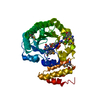

 Mass: 456.344 Da / Num. of mol.: 1 / Source method: obtained synthetically / Formula: C17H21N4O9P
 Mass: 18.015 Da / Num. of mol.: 104 / Source method: isolated from a natural source / Formula: H2O
 Sample preparation
 / Beamline: I04 / Wavelength: 0.9808
  Processing