 Movie
Movie Controller


+ Open data

- Basic information
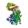
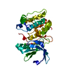
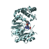
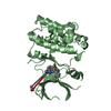
| Entry | Database: PDB / ID: 4bf2 | ||||||
|---|---|---|---|---|---|---|---|
| Title | Crystal Structures of Ask1-inhibitor Complexes | ||||||
 Components Components | MITOGEN-ACTIVATED PROTEIN KINASE KINASE KINASE 5 | ||||||
 Keywords Keywords | TRANSFERASE | ||||||
| Function / homology |  Function and homology information Function and homology informationcellular response to reactive nitrogen species / neuron intrinsic apoptotic signaling pathway in response to oxidative stress / IRE1-TRAF2-ASK1 complex / protein kinase complex / mitogen-activated protein kinase kinase kinase / programmed necrotic cell death / JUN kinase kinase kinase activity / endothelial cell apoptotic process / positive regulation of p38MAPK cascade / intrinsic apoptotic signaling pathway in response to oxidative stress ...cellular response to reactive nitrogen species / neuron intrinsic apoptotic signaling pathway in response to oxidative stress / IRE1-TRAF2-ASK1 complex / protein kinase complex / mitogen-activated protein kinase kinase kinase / programmed necrotic cell death / JUN kinase kinase kinase activity / endothelial cell apoptotic process / positive regulation of p38MAPK cascade / intrinsic apoptotic signaling pathway in response to oxidative stress / p38MAPK cascade / intrinsic apoptotic signaling pathway in response to endoplasmic reticulum stress / MAP kinase kinase kinase activity / positive regulation of myoblast differentiation / stress-activated MAPK cascade / positive regulation of vascular associated smooth muscle cell proliferation / JNK cascade / positive regulation of cardiac muscle cell apoptotic process / response to endoplasmic reticulum stress / response to ischemia / cellular response to amino acid starvation / apoptotic signaling pathway / positive regulation of JNK cascade / cellular response to tumor necrosis factor / cellular response to hydrogen peroxide / cellular senescence / MAPK cascade / neuron apoptotic process / protein phosphatase binding / Oxidative Stress Induced Senescence / protein kinase activity / positive regulation of apoptotic process / external side of plasma membrane / protein domain specific binding / innate immune response / protein serine kinase activity / protein serine/threonine kinase activity / protein kinase binding / positive regulation of DNA-templated transcription / magnesium ion binding / protein homodimerization activity / protein-containing complex / ATP binding / identical protein binding / cytoplasm / cytosol Similarity search - Function | ||||||
| Biological species |  HOMO SAPIENS (human) HOMO SAPIENS (human) | ||||||
| Method |  X-RAY DIFFRACTION / SYNCHROTRON / MOLECULAR REPLACEMENT / Resolution: 2.11 Å X-RAY DIFFRACTION / SYNCHROTRON / MOLECULAR REPLACEMENT / Resolution: 2.11 Å | ||||||
 Authors Authors | Singh, O. / Shillings, A. / Craggs, P. / Wall, I. / Rowland, P. / Skarzynski, T. / Hobbs, C.I. / Hardwick, P. / Tanner, R. / Blunt, M. ...Singh, O. / Shillings, A. / Craggs, P. / Wall, I. / Rowland, P. / Skarzynski, T. / Hobbs, C.I. / Hardwick, P. / Tanner, R. / Blunt, M. / Witty, D.R. / Smith, K.J. | ||||||
 Citation Citation | Journal: Protein Sci. / Year: 2013 Title: Crystal Structures of Ask1-Inhibtor Complexes Provide a Platform for Structure Based Drug Design. Authors: Singh, O. / Shillings, A. / Craggs, P. / Wall, I. / Rowland, P. / Skarzynski, T. / Hobbs, C.I. / Hardwick, P. / Tanner, R. / Blunt, M. / Witty, D.R. / Smith, K.J. | ||||||
| History |
|
- Structure visualization
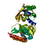
| Structure viewer | Molecule: MolmilJmol/JSmol |
|---|
- Downloads & links
-Download
| PDBx/mmCIF format | 4bf2.cif.gz | 121.2 KB | Display | PDBx/mmCIF format |
|---|---|---|---|---|
| PDB format | pdb4bf2.ent.gz | 91.9 KB | Display | PDB format |
| PDBx/mmJSON format | 4bf2.json.gz | Tree view | PDBx/mmJSON format | |
| Others |  Other downloads Other downloads |
-Validation report
| Arichive directory | https://data.pdbj.org/pub/pdb/validation_reports/bf/4bf2ftp://data.pdbj.org/pub/pdb/validation_reports/bf/4bf2 | HTTPS FTP |
|---|
-Related structure data
| Related structure data | 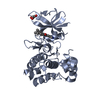 4bhnC 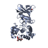 4bibC 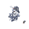 4bicC 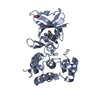 4bidC 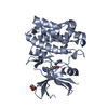 4bieC 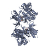 2clqS C: citing same article ( S: Starting model for refinement |
|---|---|
| Similar structure data |


-Links
 PDBj
PDBj- Assembly
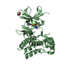
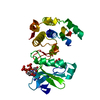
| Deposited unit | 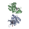
| ||||||||
|---|---|---|---|---|---|---|---|---|---|
| 1 | 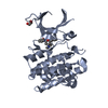
| ||||||||
| 2 | 
| ||||||||
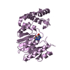
| Unit cell |
|
-Components
| #1: Protein | Mass: 37518.453 Da / Num. of mol.: 2 / Fragment: KINASE DOMAIN, RESIDUES 660-977 / Mutation: YES Source method: isolated from a genetically manipulated source Source: (gene. exp.) HOMO SAPIENS (human) / Plasmid: PFASTBAC1 / Cell line (production host): SF9 / Production host:   SPODOPTERA FRUGIPERDA (fall armyworm) SPODOPTERA FRUGIPERDA (fall armyworm)References: UniProt: Q99683, mitogen-activated protein kinase kinase #2: Chemical | 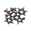  Mass: 466.531 Da / Num. of mol.: 2 / Source method: obtained synthetically / Formula: C28H26N4O3 / Comment: anticancer, antifungal, antibiotic, alkaloid*YM Mass: 466.531 Da / Num. of mol.: 2 / Source method: obtained synthetically / Formula: C28H26N4O3 / Comment: anticancer, antifungal, antibiotic, alkaloid*YM#3: Chemical | ChemComp-GOL / |   Mass: 92.094 Da / Num. of mol.: 1 / Source method: obtained synthetically / Formula: C3H8O3 Mass: 92.094 Da / Num. of mol.: 1 / Source method: obtained synthetically / Formula: C3H8O3#4: Chemical | ChemComp-ACT / |   Mass: 59.044 Da / Num. of mol.: 1 / Source method: obtained synthetically / Formula: C2H3O2 Mass: 59.044 Da / Num. of mol.: 1 / Source method: obtained synthetically / Formula: C2H3O2#5: Water | ChemComp-HOH / |  Mass: 18.015 Da / Num. of mol.: 220 / Source method: isolated from a natural source / Formula: H2O Mass: 18.015 Da / Num. of mol.: 220 / Source method: isolated from a natural source / Formula: H2O |
|---|
-Experimental details
-Experiment
| Experiment | Method: X-RAY DIFFRACTION / Number of used crystals: 1 |
|---|
- Sample preparation
| Crystal | Density Matthews: 2.48 Å3/Da / Density % sol: 50.43 % / Description: NONE |
|---|---|
| Crystal grow | pH: 6.5 Details: 18% PEG 3.4K, 0.2M NA ACETATE, 0.1M BIS-TRIS BUFFER PH 6.5, 0.2% ISOPROPANOL |
-Data collection
| Diffraction | Mean temperature: 100 K |
|---|---|
| Diffraction source | Source: SYNCHROTRON / Site: ESRF  / Beamline: ID23-1 / Wavelength: 1.072 / Beamline: ID23-1 / Wavelength: 1.072 |
| Detector | Type: ADSC CCD / Detector: CCD / Date: May 1, 2008 |
| Radiation | Protocol: SINGLE WAVELENGTH / Monochromatic (M) / Laue (L): M / Scattering type: x-ray |
| Radiation wavelength | Wavelength: 1.072 Å / Relative weight: 1 |
| Reflection | Resolution: 2.11→40 Å / Num. obs: 42333 / % possible obs: 92.7 % / Observed criterion σ(I): 2 / Redundancy: 4 % / Biso Wilson estimate: 48.3 Å2 / Rmerge(I) obs: 0.08 / Net I/σ(I): 18.3 |
| Reflection shell | Resolution: 2.11→2.15 Å / Redundancy: 4.3 % / Rmerge(I) obs: 0.51 / Mean I/σ(I) obs: 2.2 / % possible all: 97.8 |
- Processing
| Software |
| ||||||||||||||||||||||||||||||||||||||||||||||||||||||||||||||||||||||||||||||||||||||||||||||||||||||||||||||||||
|---|---|---|---|---|---|---|---|---|---|---|---|---|---|---|---|---|---|---|---|---|---|---|---|---|---|---|---|---|---|---|---|---|---|---|---|---|---|---|---|---|---|---|---|---|---|---|---|---|---|---|---|---|---|---|---|---|---|---|---|---|---|---|---|---|---|---|---|---|---|---|---|---|---|---|---|---|---|---|---|---|---|---|---|---|---|---|---|---|---|---|---|---|---|---|---|---|---|---|---|---|---|---|---|---|---|---|---|---|---|---|---|---|---|---|---|
| Refinement | Method to determine structure: MOLECULAR REPLACEMENT Starting model: PDB ENTRY 2CLQ Resolution: 2.11→13.36 Å / Cor.coef. Fo:Fc: 0.9405 / Cor.coef. Fo:Fc free: 0.9142 / SU R Cruickshank DPI: 0.181 / Cross valid method: THROUGHOUT / σ(F): 0 / SU R Blow DPI: 0.19 / SU Rfree Blow DPI: 0.172 / SU Rfree Cruickshank DPI: 0.168
| ||||||||||||||||||||||||||||||||||||||||||||||||||||||||||||||||||||||||||||||||||||||||||||||||||||||||||||||||||
| Displacement parameters | Biso mean: 53.14 Å2
| ||||||||||||||||||||||||||||||||||||||||||||||||||||||||||||||||||||||||||||||||||||||||||||||||||||||||||||||||||
| Refine analyze | Luzzati coordinate error obs: 0.29 Å | ||||||||||||||||||||||||||||||||||||||||||||||||||||||||||||||||||||||||||||||||||||||||||||||||||||||||||||||||||
| Refinement step | Cycle: LAST / Resolution: 2.11→13.36 Å
| ||||||||||||||||||||||||||||||||||||||||||||||||||||||||||||||||||||||||||||||||||||||||||||||||||||||||||||||||||
| Refine LS restraints |
| ||||||||||||||||||||||||||||||||||||||||||||||||||||||||||||||||||||||||||||||||||||||||||||||||||||||||||||||||||
| LS refinement shell | Resolution: 2.11→2.17 Å / Total num. of bins used: 20
|
