 Movie
Movie Controller


+ Open data

- Basic information
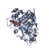
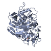
| Entry | Database: PDB / ID: 4bbo | ||||||
|---|---|---|---|---|---|---|---|
| Title | Crystal structure of core-bradavidin | ||||||
 Components Components | BLR5658 PROTEIN | ||||||
 Keywords Keywords | BIOTIN-BINDING PROTEIN / AVIDINS | ||||||
| Function / homology |  Function and homology information Function and homology information | ||||||
| Biological species |  BRADYRHIZOBIUM JAPONICUM (bacteria) BRADYRHIZOBIUM JAPONICUM (bacteria) | ||||||
| Method |  X-RAY DIFFRACTION / SYNCHROTRON / MOLECULAR REPLACEMENT / Resolution: 1.6 Å X-RAY DIFFRACTION / SYNCHROTRON / MOLECULAR REPLACEMENT / Resolution: 1.6 Å | ||||||
 Authors Authors | Airenne, T.T. / Johnson, M.S. / Maatta, J.A.E. / Hytonen, V.H. / Kulomaa, M.S. | ||||||
 Citation Citation | Journal: To be Published Title: Crystal Structure of Core-Bradavidin Authors: Airenne, T.T. / Maatta, J.A.E. / Nordlund, H. / Kulomaa, M.S. / Johnson, M.S. | ||||||
| History |
| ||||||
| Remark 700 | SHEET DETERMINATION METHOD: DSSP THE SHEETS PRESENTED AS "AA" IN EACH CHAIN ON SHEET RECORDS BELOW ... SHEET DETERMINATION METHOD: DSSP THE SHEETS PRESENTED AS "AA" IN EACH CHAIN ON SHEET RECORDS BELOW IS ACTUALLY AN 8-STRANDED BARREL THIS IS REPRESENTED BY A 9-STRANDED SHEET IN WHICH THE FIRST AND LAST STRANDS ARE IDENTICAL. THE SHEETS PRESENTED AS "BA" IN EACH CHAIN ON SHEET RECORDS BELOW IS ACTUALLY AN 8-STRANDED BARREL THIS IS REPRESENTED BY A 9-STRANDED SHEET IN WHICH THE FIRST AND LAST STRANDS ARE IDENTICAL. THE SHEETS PRESENTED AS "CA" IN EACH CHAIN ON SHEET RECORDS BELOW IS ACTUALLY AN 8-STRANDED BARREL THIS IS REPRESENTED BY A 9-STRANDED SHEET IN WHICH THE FIRST AND LAST STRANDS ARE IDENTICAL. THE SHEETS PRESENTED AS "DA" IN EACH CHAIN ON SHEET RECORDS BELOW IS ACTUALLY AN 8-STRANDED BARREL THIS IS REPRESENTED BY A 9-STRANDED SHEET IN WHICH THE FIRST AND LAST STRANDS ARE IDENTICAL. |
- Structure visualization
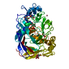
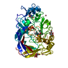
| Structure viewer | Molecule: MolmilJmol/JSmol |
|---|
- Downloads & links
-Download
| PDBx/mmCIF format | 4bbo.cif.gz | 182.7 KB | Display | PDBx/mmCIF format |
|---|---|---|---|---|
| PDB format | pdb4bbo.ent.gz | 148 KB | Display | PDB format |
| PDBx/mmJSON format | 4bbo.json.gz | Tree view | PDBx/mmJSON format | |
| Others |  Other downloads Other downloads |
-Validation report
| Arichive directory | https://data.pdbj.org/pub/pdb/validation_reports/bb/4bboftp://data.pdbj.org/pub/pdb/validation_reports/bb/4bbo | HTTPS FTP |
|---|
-Related structure data
| Related structure data |  2y32S S: Starting model for refinement |
|---|---|
| Similar structure data |


-Links
 PDBj
PDBj- Assembly
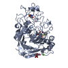
| Deposited unit | 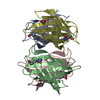
| ||||||||||||||||||||||||||||
|---|---|---|---|---|---|---|---|---|---|---|---|---|---|---|---|---|---|---|---|---|---|---|---|---|---|---|---|---|---|
| 1 |
| ||||||||||||||||||||||||||||
| Unit cell |
| ||||||||||||||||||||||||||||
| Noncrystallographic symmetry (NCS) | NCS oper:
|
-Components
| #1: Protein | Mass: 12300.226 Da / Num. of mol.: 4 / Fragment: RESIDUES 26-143 Source method: isolated from a genetically manipulated source Source: (gene. exp.) BRADYRHIZOBIUM JAPONICUM (bacteria) / Production host: #2: Chemical | ChemComp-BTN /   Mass: 244.311 Da / Num. of mol.: 4 / Source method: obtained synthetically / Formula: C10H16N2O3S Mass: 244.311 Da / Num. of mol.: 4 / Source method: obtained synthetically / Formula: C10H16N2O3S#3: Chemical | ChemComp-GOL / |   Mass: 92.094 Da / Num. of mol.: 1 / Source method: obtained synthetically / Formula: C3H8O3 Mass: 92.094 Da / Num. of mol.: 1 / Source method: obtained synthetically / Formula: C3H8O3#4: Chemical |   Mass: 59.044 Da / Num. of mol.: 3 / Source method: obtained synthetically / Formula: C2H3O2 Mass: 59.044 Da / Num. of mol.: 3 / Source method: obtained synthetically / Formula: C2H3O2#5: Water | ChemComp-HOH / |  Mass: 18.015 Da / Num. of mol.: 368 / Source method: isolated from a natural source / Formula: H2O Mass: 18.015 Da / Num. of mol.: 368 / Source method: isolated from a natural source / Formula: H2OHas protein modification | Y | |
|---|
-Experimental details
-Experiment
| Experiment | Method: X-RAY DIFFRACTION / Number of used crystals: 1 |
|---|
- Sample preparation
| Crystal | Density Matthews: 2 Å3/Da / Density % sol: 38.68 % Description: CORE REGION OF 2Y32 WAS USED FOR MOLECULAR REPLACEMENT. |
|---|---|
| Crystal grow | Method: vapor diffusion, hanging drop Details: ONE MICROLITER OF THE PROTEIN SOLUTION (LESS THAN 1 MG PER ML AND IN 50 MM SODIUM ACETATE, 100 MM NACL, PH 4) WAS CRYSTALLIZED BY ADDING ONE MICROLITER OF THE WELL SOLUTION (0.1 M HEPES PH 7. ...Details: ONE MICROLITER OF THE PROTEIN SOLUTION (LESS THAN 1 MG PER ML AND IN 50 MM SODIUM ACETATE, 100 MM NACL, PH 4) WAS CRYSTALLIZED BY ADDING ONE MICROLITER OF THE WELL SOLUTION (0.1 M HEPES PH 7.4, 0.8 M K,NA TARTRATE).HANGING DROPS AND VAPOUR DIFFUSION METHOD WAS USED AT RT. BEFORE CRYSTALLIZATION, 25 MICROLITERS OF THE PROTEIN SAMPLE WAS INCUBATED WITH 1 MICRO LITER OF BIOTIN SOLUTION (1 MG PER ML AND IN 5 MM TRIS PH 8.8, 8 MM CHES PH 9.5) FOR 3.5 HOURS AT 37 DEGREES OF CELCIUS. |
-Data collection
| Diffraction | Mean temperature: 100 K |
|---|---|
| Diffraction source | Source: SYNCHROTRON / Site: MAX II  / Beamline: I711 / Wavelength: 1.063 / Beamline: I711 / Wavelength: 1.063 |
| Detector | Date: Apr 21, 2005 |
| Radiation | Protocol: SINGLE WAVELENGTH / Monochromatic (M) / Laue (L): M / Scattering type: x-ray |
| Radiation wavelength | Wavelength: 1.063 Å / Relative weight: 1 |
| Reflection | Resolution: 1.6→25 Å / Num. obs: 52798 / % possible obs: 100 % / Observed criterion σ(I): -3 / Redundancy: 9.6 % / Rmerge(I) obs: 0.09 / Net I/σ(I): 18 |
| Reflection shell | Resolution: 1.6→1.7 Å / Redundancy: 9.6 % / Rmerge(I) obs: 0.55 / Mean I/σ(I) obs: 4 / % possible all: 100 |
- Processing
| Software |
| ||||||||||||||||||||||||||||||||||||||||||||||||||||||||||||||||||||||||||||||||||||||||||||||||||||||||||||||||||||||||||||||||||||||||||||||||||||||||||||||||||||||||||||||||||||||
|---|---|---|---|---|---|---|---|---|---|---|---|---|---|---|---|---|---|---|---|---|---|---|---|---|---|---|---|---|---|---|---|---|---|---|---|---|---|---|---|---|---|---|---|---|---|---|---|---|---|---|---|---|---|---|---|---|---|---|---|---|---|---|---|---|---|---|---|---|---|---|---|---|---|---|---|---|---|---|---|---|---|---|---|---|---|---|---|---|---|---|---|---|---|---|---|---|---|---|---|---|---|---|---|---|---|---|---|---|---|---|---|---|---|---|---|---|---|---|---|---|---|---|---|---|---|---|---|---|---|---|---|---|---|---|---|---|---|---|---|---|---|---|---|---|---|---|---|---|---|---|---|---|---|---|---|---|---|---|---|---|---|---|---|---|---|---|---|---|---|---|---|---|---|---|---|---|---|---|---|---|---|---|---|
| Refinement | Method to determine structure: MOLECULAR REPLACEMENT Starting model: PDB ENTRY 2Y32 Resolution: 1.6→61.9 Å / Cor.coef. Fo:Fc: 0.971 / Cor.coef. Fo:Fc free: 0.96 / SU B: 2.959 / SU ML: 0.05 / Cross valid method: THROUGHOUT / ESU R: 0.079 / ESU R Free: 0.08 / Stereochemistry target values: MAXIMUM LIKELIHOOD / Details: HYDROGENS HAVE BEEN ADDED IN THE RIDING POSITIONS.
| ||||||||||||||||||||||||||||||||||||||||||||||||||||||||||||||||||||||||||||||||||||||||||||||||||||||||||||||||||||||||||||||||||||||||||||||||||||||||||||||||||||||||||||||||||||||
| Solvent computation | Ion probe radii: 0.8 Å / Shrinkage radii: 0.8 Å / VDW probe radii: 1.2 Å / Solvent model: MASK | ||||||||||||||||||||||||||||||||||||||||||||||||||||||||||||||||||||||||||||||||||||||||||||||||||||||||||||||||||||||||||||||||||||||||||||||||||||||||||||||||||||||||||||||||||||||
| Displacement parameters | Biso mean: 14.577 Å2
| ||||||||||||||||||||||||||||||||||||||||||||||||||||||||||||||||||||||||||||||||||||||||||||||||||||||||||||||||||||||||||||||||||||||||||||||||||||||||||||||||||||||||||||||||||||||
| Refinement step | Cycle: LAST / Resolution: 1.6→61.9 Å
| ||||||||||||||||||||||||||||||||||||||||||||||||||||||||||||||||||||||||||||||||||||||||||||||||||||||||||||||||||||||||||||||||||||||||||||||||||||||||||||||||||||||||||||||||||||||
| Refine LS restraints |
|