 Movie
Movie Controller

[English] 日本語
 Yorodumi
- PDB-4ba5: Crystal structure of omega-transaminase from Chromobacterium violaceum -
+ Open data

- Basic information
| Entry | Database: PDB / ID: 4ba5 | ||||||
|---|---|---|---|---|---|---|---|
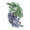
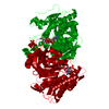
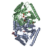
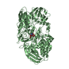
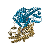
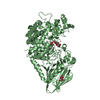
| Title | Crystal structure of omega-transaminase from Chromobacterium violaceum | ||||||
 Components Components | AMINOTRANSFERASE | ||||||
 Keywords Keywords | TRANSFERASE | ||||||
| Function / homology |  Function and homology information Function and homology informationadenosylmethionine-8-amino-7-oxononanoate transaminase / S-adenosyl-L-methionine:8-amino-7-oxononanoate transaminase activity / pyridoxal phosphate binding / identical protein binding / cytosol Similarity search - Function | ||||||
| Biological species |  CHROMOBACTERIUM VIOLACEUM (bacteria) CHROMOBACTERIUM VIOLACEUM (bacteria) | ||||||
| Method |  X-RAY DIFFRACTION / SYNCHROTRON / MOLECULAR REPLACEMENT / Resolution: 1.76 Å X-RAY DIFFRACTION / SYNCHROTRON / MOLECULAR REPLACEMENT / Resolution: 1.76 Å | ||||||
 Authors Authors | Sayer, C. / Isupov, M.N. / Littlechild, J.A. | ||||||
 Citation Citation | Journal: Acta Crystallogr.,Sect.D / Year: 2013 Title: Structural Studies with Pseudomonas and Chromobacterium [Omega]-Aminotransferases Provide Insights Into Their Differing Substrate Specificity. Authors: Sayer, C. / Isupov, M.N. / Westlake, A. / Littlechild, J.A. | ||||||
| History |
|
- Structure visualization
| Structure viewer | Molecule: MolmilJmol/JSmol |
|---|
- Downloads & links
-Download
| PDBx/mmCIF format | 4ba5.cif.gz | 198.8 KB | Display | PDBx/mmCIF format |
|---|---|---|---|---|
| PDB format | pdb4ba5.ent.gz | 159.9 KB | Display | PDB format |
| PDBx/mmJSON format | 4ba5.json.gz | Tree view | PDBx/mmJSON format | |
| Others |  Other downloads Other downloads |
-Validation report
| Arichive directory | https://data.pdbj.org/pub/pdb/validation_reports/ba/4ba5ftp://data.pdbj.org/pub/pdb/validation_reports/ba/4ba5 | HTTPS FTP |
|---|
-Related structure data
| Related structure data |  4ah3SC 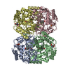 4b98C  4b9bC  4ba4C S: Starting model for refinement C: citing same article ( |
|---|---|
| Similar structure data |


-Links
 PDBj
PDBj- Assembly
| Deposited unit | 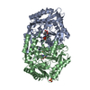
| ||||||||
|---|---|---|---|---|---|---|---|---|---|
| 1 |
| ||||||||
| Unit cell |
|
-Components
| #1: Protein | Mass: 51279.293 Da / Num. of mol.: 2 Source method: isolated from a genetically manipulated source Source: (gene. exp.) CHROMOBACTERIUM VIOLACEUM (bacteria) / Production host: References: UniProt: Q7NWG4, beta-alanine-pyruvate transaminase #2: Chemical |   Mass: 368.278 Da / Num. of mol.: 2 / Source method: obtained synthetically / Formula: C15H17N2O7P Mass: 368.278 Da / Num. of mol.: 2 / Source method: obtained synthetically / Formula: C15H17N2O7P#3: Chemical | ChemComp-SO4 / |   Mass: 96.063 Da / Num. of mol.: 1 / Source method: obtained synthetically / Formula: SO4 Mass: 96.063 Da / Num. of mol.: 1 / Source method: obtained synthetically / Formula: SO4#4: Water | ChemComp-HOH / |  Mass: 18.015 Da / Num. of mol.: 582 / Source method: isolated from a natural source / Formula: H2O Mass: 18.015 Da / Num. of mol.: 582 / Source method: isolated from a natural source / Formula: H2O |
|---|
-Experimental details
-Experiment
| Experiment | Method: X-RAY DIFFRACTION / Number of used crystals: 1 |
|---|
- Sample preparation
| Crystal | Density Matthews: 2.08 Å3/Da / Density % sol: 40.4 % / Description: NONE |
|---|
-Data collection
| Diffraction | Mean temperature: 100 K |
|---|---|
| Diffraction source | Source: SYNCHROTRON / Site: SRS  / Beamline: PX14.1 / Wavelength: 1.49 / Beamline: PX14.1 / Wavelength: 1.49 |
| Detector | Type: ADSC CCD / Detector: CCD |
| Radiation | Protocol: SINGLE WAVELENGTH / Monochromatic (M) / Laue (L): M / Scattering type: x-ray |
| Radiation wavelength | Wavelength: 1.49 Å / Relative weight: 1 |
| Reflection | Resolution: 1.73→41.7 Å / Num. obs: 65355 / % possible obs: 86.6 % / Observed criterion σ(I): 1.7 / Redundancy: 2.1 % / Rmerge(I) obs: 0.07 / Net I/σ(I): 6.8 |
| Reflection shell | Resolution: 1.73→1.82 Å / Redundancy: 2.1 % / Rmerge(I) obs: 0.31 / Mean I/σ(I) obs: 1.7 / % possible all: 52.4 |
- Processing
| Software |
| ||||||||||||||||||||
|---|---|---|---|---|---|---|---|---|---|---|---|---|---|---|---|---|---|---|---|---|---|
| Refinement | Method to determine structure: MOLECULAR REPLACEMENT Starting model: PDB ENTRY 4AH3 Resolution: 1.76→56.75 Å / Cor.coef. Fo:Fc: 0.966 / Cor.coef. Fo:Fc free: 0.94 / SU B: 3.17 / SU ML: 0.1 / Cross valid method: THROUGHOUT / ESU R: 0.151 / ESU R Free: 0.147 / Stereochemistry target values: MAXIMUM LIKELIHOOD / Details: HYDROGENS HAVE BEEN ADDED IN THE RIDING POSITIONS.
| ||||||||||||||||||||
| Displacement parameters | Biso mean: 31.192 Å2
| ||||||||||||||||||||
| Refinement step | Cycle: LAST / Resolution: 1.76→56.75 Å
| ||||||||||||||||||||
| LS refinement shell | Resolution: 1.761→1.807 Å / Total num. of bins used: 20
|
