 Movie
Movie Controller

[English] 日本語
 Yorodumi
- PDB-4b6r: Structure of Helicobacter pylori Type II Dehydroquinase inhibited... -
+ Open data

- Basic information
| Entry | Database: PDB / ID: 4b6r | ||||||
|---|---|---|---|---|---|---|---|
| Title | Structure of Helicobacter pylori Type II Dehydroquinase inhibited by (2S)-2-(4-methoxy)benzyl-3-dehydroquinic acid | ||||||
 Components Components | 3-DEHYDROQUINATE DEHYDRATASE | ||||||
 Keywords Keywords | LYASE | ||||||
| Function / homology |  Function and homology information Function and homology informationquinate catabolic process / 3-dehydroquinate dehydratase / 3-dehydroquinate dehydratase activity / chorismate biosynthetic process / aromatic amino acid biosynthetic process / amino acid biosynthetic process Similarity search - Function | ||||||
| Biological species |  HELICOBACTER PYLORI 26695 (bacteria) HELICOBACTER PYLORI 26695 (bacteria) | ||||||
| Method |  X-RAY DIFFRACTION / SYNCHROTRON / MOLECULAR REPLACEMENT / Resolution: 2 Å X-RAY DIFFRACTION / SYNCHROTRON / MOLECULAR REPLACEMENT / Resolution: 2 Å | ||||||
 Authors Authors | Otero, J.M. / Llamas-Saiz, A.L. / Lence, E. / Tizon, L. / Peon, A. / Prazeres, V.F.V. / Lamb, H. / Hawkins, A.R. / Gonzalez-Bello, C. / van Raaij, M.J. | ||||||
 Citation Citation | Journal: ACS Chem. Biol. / Year: 2013 Title: Mechanistic basis of the inhibition of type II dehydroquinase by (2S)- and (2R)-2-benzyl-3-dehydroquinic acids. Authors: Lence, E. / Tizon, L. / Otero, J.M. / Peon, A. / Prazeres, V.F. / Llamas-Saiz, A.L. / Fox, G.C. / van Raaij, M.J. / Lamb, H. / Hawkins, A.R. / Gonzalez-Bello, C. | ||||||
| History |
|
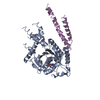
- Structure visualization
| Structure viewer | Molecule: MolmilJmol/JSmol |
|---|
- Downloads & links
-Download
| PDBx/mmCIF format | 4b6r.cif.gz | 109.9 KB | Display | PDBx/mmCIF format |
|---|---|---|---|---|
| PDB format | pdb4b6r.ent.gz | 85.8 KB | Display | PDB format |
| PDBx/mmJSON format | 4b6r.json.gz | Tree view | PDBx/mmJSON format | |
| Others |  Other downloads Other downloads |
-Validation report
| Arichive directory | https://data.pdbj.org/pub/pdb/validation_reports/b6/4b6rftp://data.pdbj.org/pub/pdb/validation_reports/b6/4b6r | HTTPS FTP |
|---|
-Related structure data
| Related structure data |  4b6oC  4b6pC  4b6qC  4b6sC  2c4vS S: Starting model for refinement C: citing same article ( |
|---|---|
| Similar structure data |


-Links
 PDBj
PDBj
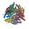
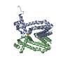
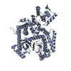
- Assembly
| Deposited unit | 
| ||||||||
|---|---|---|---|---|---|---|---|---|---|
| 1 |
| ||||||||
| Unit cell |
|
-Components
| #1: Protein | Mass: 18499.246 Da / Num. of mol.: 3 Source method: isolated from a genetically manipulated source Source: (gene. exp.) HELICOBACTER PYLORI 26695 (bacteria) / Plasmid: PET21A / Production host: #2: Chemical | 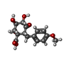  Mass: 310.299 Da / Num. of mol.: 3 / Source method: obtained synthetically / Formula: C15H18O7 Mass: 310.299 Da / Num. of mol.: 3 / Source method: obtained synthetically / Formula: C15H18O7#3: Chemical | ChemComp-SO4 / |   Mass: 96.063 Da / Num. of mol.: 1 / Source method: obtained synthetically / Formula: SO4 Mass: 96.063 Da / Num. of mol.: 1 / Source method: obtained synthetically / Formula: SO4#4: Water | ChemComp-HOH / |  Mass: 18.015 Da / Num. of mol.: 179 / Source method: isolated from a natural source / Formula: H2O Mass: 18.015 Da / Num. of mol.: 179 / Source method: isolated from a natural source / Formula: H2O |
|---|
-Experimental details
-Experiment
| Experiment | Method: X-RAY DIFFRACTION / Number of used crystals: 1 |
|---|
- Sample preparation
| Crystal | Density Matthews: 2.4 Å3/Da / Density % sol: 48 % / Description: NONE |
|---|---|
| Crystal grow | pH: 5.4 Details: 50 MM TRIS-HCL PH 7.5, 1 MM 2-MERCAPTOETHANOL, 1 MM ETHYLENEDIAMINETETRAACETIC ACID, 200 MM SODIUM CHLORIDE 36% (W/V) POLYETHYLENEGLYCOL 4000, 0.1 M SODIUM CITRATE PH 5.4 |
-Data collection
| Diffraction | Mean temperature: 100 K |
|---|---|
| Diffraction source | Source: SYNCHROTRON / Site: SOLEIL  / Beamline: PROXIMA 1 / Wavelength: 0.98011 / Beamline: PROXIMA 1 / Wavelength: 0.98011 |
| Detector | Type: ADSC QUANTUM 315r / Detector: CCD / Date: Jul 26, 2010 / Details: KIRKPATRICK-BAEZ PAIR OF BI-MORPH MIRRORS |
| Radiation | Monochromator: CHANNEL CUT CRYOGENICALLY COOLED MONOCHROMATOR CRYSTAL Protocol: SINGLE WAVELENGTH / Monochromatic (M) / Laue (L): M / Scattering type: x-ray |
| Radiation wavelength | Wavelength: 0.98011 Å / Relative weight: 1 |
| Reflection | Resolution: 2→58.7 Å / Num. obs: 36522 / % possible obs: 99.7 % / Redundancy: 4.4 % / Biso Wilson estimate: 33.8 Å2 / Rmerge(I) obs: 0.08 / Net I/σ(I): 4.9 |
| Reflection shell | Resolution: 2→2.11 Å / Redundancy: 3.9 % / Rmerge(I) obs: 0.34 / Mean I/σ(I) obs: 2.2 / % possible all: 100 |
- Processing
| Software |
| ||||||||||||||||||||||||||||||||||||||||||||||||||||||||||||||||||||||||||||||||||||||||||||||||||||||||||||||||||||||||||||||||||||||||||||||||||||||||||||||||||||||||||||||||||||||
|---|---|---|---|---|---|---|---|---|---|---|---|---|---|---|---|---|---|---|---|---|---|---|---|---|---|---|---|---|---|---|---|---|---|---|---|---|---|---|---|---|---|---|---|---|---|---|---|---|---|---|---|---|---|---|---|---|---|---|---|---|---|---|---|---|---|---|---|---|---|---|---|---|---|---|---|---|---|---|---|---|---|---|---|---|---|---|---|---|---|---|---|---|---|---|---|---|---|---|---|---|---|---|---|---|---|---|---|---|---|---|---|---|---|---|---|---|---|---|---|---|---|---|---|---|---|---|---|---|---|---|---|---|---|---|---|---|---|---|---|---|---|---|---|---|---|---|---|---|---|---|---|---|---|---|---|---|---|---|---|---|---|---|---|---|---|---|---|---|---|---|---|---|---|---|---|---|---|---|---|---|---|---|---|
| Refinement | Method to determine structure: MOLECULAR REPLACEMENT Starting model: PDB ENTRY 2C4V Resolution: 2→58.7 Å / Cor.coef. Fo:Fc: 0.953 / Cor.coef. Fo:Fc free: 0.888 / SU B: 5.176 / SU ML: 0.144 / Cross valid method: THROUGHOUT / ESU R: 0.198 / ESU R Free: 0.19 / Stereochemistry target values: MAXIMUM LIKELIHOOD Details: HYDROGENS HAVE BEEN ADDED IN THE RIDING POSITIONS. U VALUES REFINED INDIVIDUALLY
| ||||||||||||||||||||||||||||||||||||||||||||||||||||||||||||||||||||||||||||||||||||||||||||||||||||||||||||||||||||||||||||||||||||||||||||||||||||||||||||||||||||||||||||||||||||||
| Solvent computation | Ion probe radii: 0.8 Å / Shrinkage radii: 0.8 Å / VDW probe radii: 1.4 Å / Solvent model: MASK | ||||||||||||||||||||||||||||||||||||||||||||||||||||||||||||||||||||||||||||||||||||||||||||||||||||||||||||||||||||||||||||||||||||||||||||||||||||||||||||||||||||||||||||||||||||||
| Displacement parameters | Biso mean: 45.424 Å2
| ||||||||||||||||||||||||||||||||||||||||||||||||||||||||||||||||||||||||||||||||||||||||||||||||||||||||||||||||||||||||||||||||||||||||||||||||||||||||||||||||||||||||||||||||||||||
| Refinement step | Cycle: LAST / Resolution: 2→58.7 Å
| ||||||||||||||||||||||||||||||||||||||||||||||||||||||||||||||||||||||||||||||||||||||||||||||||||||||||||||||||||||||||||||||||||||||||||||||||||||||||||||||||||||||||||||||||||||||
| Refine LS restraints |
|