 Movie
Movie Controller


+ Open data

- Basic information
| Entry | Database: PDB / ID: 1j2y | ||||||
|---|---|---|---|---|---|---|---|
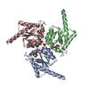
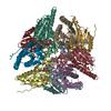
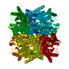
| Title | Crystal structure of the type II 3-dehydroquinase | ||||||
 Components Components | 3-dehydroquinate dehydratase | ||||||
 Keywords Keywords | LYASE / 3-dehydroquinase | ||||||
| Function / homology |  Function and homology information Function and homology informationquinate catabolic process / 3-dehydroquinate dehydratase / 3-dehydroquinate dehydratase activity / chorismate biosynthetic process / aromatic amino acid biosynthetic process / amino acid biosynthetic process Similarity search - Function | ||||||
| Biological species |   Helicobacter pylori (bacteria) Helicobacter pylori (bacteria) | ||||||
| Method |  X-RAY DIFFRACTION / SYNCHROTRON / MOLECULAR REPLACEMENT / Resolution: 2.6 Å X-RAY DIFFRACTION / SYNCHROTRON / MOLECULAR REPLACEMENT / Resolution: 2.6 Å | ||||||
 Authors Authors | Lee, B.I. / Kwak, J.E. / Suh, S.W. | ||||||
 Citation Citation | Journal: Proteins / Year: 2003 Title: Crystal structure of the type II 3-dehydroquinase from Helicobacter pylori Authors: Lee, B.I. / Kwak, J.E. / Suh, S.W. | ||||||
| History |
|
- Structure visualization
| Structure viewer | Molecule: MolmilJmol/JSmol |
|---|
- Downloads & links
-Download
| PDBx/mmCIF format | 1j2y.cif.gz | 44.7 KB | Display | PDBx/mmCIF format |
|---|---|---|---|---|
| PDB format | pdb1j2y.ent.gz | 31.5 KB | Display | PDB format |
| PDBx/mmJSON format | 1j2y.json.gz | Tree view | PDBx/mmJSON format | |
| Others |  Other downloads Other downloads |
-Validation report
| Arichive directory | https://data.pdbj.org/pub/pdb/validation_reports/j2/1j2yftp://data.pdbj.org/pub/pdb/validation_reports/j2/1j2y | HTTPS FTP |
|---|
-Related structure data
| Related structure data |  2dhqS S: Starting model for refinement |
|---|---|
| Similar structure data |


-Links
 PDBj
PDBj
- Assembly
| Deposited unit | 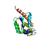
| ||||||||
|---|---|---|---|---|---|---|---|---|---|
| 1 | x 12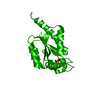
| ||||||||
| Unit cell |
| ||||||||
| Details | The biological assembly is dodecamer generated from the monomer in the asymmetric unit by the operations: z, x-1, y+1 and y+1,z-1,x and -x+2, y, -z+2 and -y+1, z-1, -x+2 and -z+2,x-1,-y+1 and -z+2, -x+1, y+1 and -y+1,-z+1,x and -x+2,-y,z and y+1, -z+1, -x+2 and x, -y, -z+2 and z,-x+1, -y+1 and |
-Components
| #1: Protein | Mass: 18499.246 Da / Num. of mol.: 1 Source method: isolated from a genetically manipulated source Source: (gene. exp.) Helicobacter pylori (bacteria) / Gene: aroQ / Plasmid: pET21a / Production host: |
|---|---|
| #2: Chemical | ChemComp-DQA /   Mass: 190.151 Da / Num. of mol.: 1 / Source method: obtained synthetically / Formula: C7H10O6 Mass: 190.151 Da / Num. of mol.: 1 / Source method: obtained synthetically / Formula: C7H10O6 |
| #3: Water | ChemComp-HOH /  Mass: 18.015 Da / Num. of mol.: 19 / Source method: isolated from a natural source / Formula: H2O Mass: 18.015 Da / Num. of mol.: 19 / Source method: isolated from a natural source / Formula: H2O |
-Experimental details
-Experiment
| Experiment | Method: X-RAY DIFFRACTION / Number of used crystals: 1 |
|---|
- Sample preparation
| Crystal | Density Matthews: 2.3 Å3/Da / Density % sol: 46.07 % |
|---|---|
| Crystal grow | Temperature: 295 K / Method: vapor diffusion, hanging drop / pH: 5.8 Details: PEG 4000, sodium cholride, sodium citrate, EDTA, 2-mercaptoethanol, pH 5.8, VAPOR DIFFUSION, HANGING DROP, temperature 295K |
| Crystal grow | *PLUS Temperature: 296 K / Method: vapor diffusion, hanging drop / Details: Kwak, J.E., (2001) Acta Crystallogr., D57, 279. |
| Components of the solutions | *PLUS Conc.: 20 mg/ml / Common name: protein |
-Data collection
| Diffraction | Mean temperature: 100 K |
|---|---|
| Diffraction source | Source: SYNCHROTRON / Site: Photon Factory  / Beamline: BL-6A / Wavelength: 1 Å / Beamline: BL-6A / Wavelength: 1 Å |
| Detector | Type: ADSC QUANTUM 4 / Detector: CCD |
| Radiation | Protocol: SINGLE WAVELENGTH / Monochromatic (M) / Laue (L): M / Scattering type: x-ray |
| Radiation wavelength | Wavelength: 1 Å / Relative weight: 1 |
| Reflection | Resolution: 2.5→44 Å / Num. all: 6146 / Num. obs: 6146 / % possible obs: 100 % / Observed criterion σ(F): 0 / Observed criterion σ(I): 0 / Redundancy: 38 % / Biso Wilson estimate: 53.5 Å2 / Rsym value: 0.086 |
| Reflection shell | Resolution: 2.5→2.64 Å / Rsym value: 0.484 / % possible all: 100 |
- Processing
| Software |
| ||||||||||||||||||||||||||||||||||||||||||||||||||||||||||||||||||||||||||||||||
|---|---|---|---|---|---|---|---|---|---|---|---|---|---|---|---|---|---|---|---|---|---|---|---|---|---|---|---|---|---|---|---|---|---|---|---|---|---|---|---|---|---|---|---|---|---|---|---|---|---|---|---|---|---|---|---|---|---|---|---|---|---|---|---|---|---|---|---|---|---|---|---|---|---|---|---|---|---|---|---|---|---|
| Refinement | Method to determine structure: MOLECULAR REPLACEMENT Starting model: PDB ENTRY 2DHQ Resolution: 2.6→19.78 Å / Rfactor Rfree error: 0.012 / Data cutoff high absF: 160671.11 / Data cutoff low absF: 0 / Isotropic thermal model: RESTRAINED / Cross valid method: THROUGHOUT / σ(F): 2
| ||||||||||||||||||||||||||||||||||||||||||||||||||||||||||||||||||||||||||||||||
| Solvent computation | Solvent model: FLAT MODEL / Bsol: 46.0261 Å2 / ksol: 0.311869 e/Å3 | ||||||||||||||||||||||||||||||||||||||||||||||||||||||||||||||||||||||||||||||||
| Displacement parameters | Biso mean: 52.2 Å2
| ||||||||||||||||||||||||||||||||||||||||||||||||||||||||||||||||||||||||||||||||
| Refine analyze |
| ||||||||||||||||||||||||||||||||||||||||||||||||||||||||||||||||||||||||||||||||
| Refinement step | Cycle: LAST / Resolution: 2.6→19.78 Å
| ||||||||||||||||||||||||||||||||||||||||||||||||||||||||||||||||||||||||||||||||
| Refine LS restraints |
| ||||||||||||||||||||||||||||||||||||||||||||||||||||||||||||||||||||||||||||||||
| LS refinement shell | Resolution: 2.6→2.76 Å / Rfactor Rfree error: 0.045 / Total num. of bins used: 6
| ||||||||||||||||||||||||||||||||||||||||||||||||||||||||||||||||||||||||||||||||
| Xplor file |
| ||||||||||||||||||||||||||||||||||||||||||||||||||||||||||||||||||||||||||||||||
| Refinement | *PLUS Highest resolution: 2.6 Å / Lowest resolution: 20 Å / % reflection Rfree: 10 % | ||||||||||||||||||||||||||||||||||||||||||||||||||||||||||||||||||||||||||||||||
| Solvent computation | *PLUS | ||||||||||||||||||||||||||||||||||||||||||||||||||||||||||||||||||||||||||||||||
| Displacement parameters | *PLUS | ||||||||||||||||||||||||||||||||||||||||||||||||||||||||||||||||||||||||||||||||
| Refine LS restraints | *PLUS
| ||||||||||||||||||||||||||||||||||||||||||||||||||||||||||||||||||||||||||||||||
| LS refinement shell | *PLUS Highest resolution: 2.6 Å |
