 Movie
Movie Controller

[English] 日本語
 Yorodumi
- PDB-4b0n: Crystal structure of PKS-I from the brown algae Ectocarpus siliculosus -
+ Open data

- Basic information
| Entry | Database: PDB / ID: 4b0n | ||||||
|---|---|---|---|---|---|---|---|
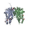
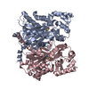
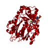
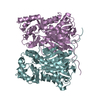
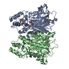
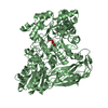
| Title | Crystal structure of PKS-I from the brown algae Ectocarpus siliculosus | ||||||
 Components Components | POLYKETIDE SYNTHASE III | ||||||
 Keywords Keywords | TRANSFERASE / POLYPHENOL BIOSYNTHESIS / ARACHIDONYL GROUP | ||||||
| Function / homology |  Function and homology information Function and homology informationpolyketide biosynthetic process / acyltransferase activity, transferring groups other than amino-acyl groups / Transferases; Acyltransferases; Transferring groups other than aminoacyl groups Similarity search - Function | ||||||
| Biological species |  ECTOCARPUS SILICULOSUS (eukaryote) ECTOCARPUS SILICULOSUS (eukaryote) | ||||||
| Method |  X-RAY DIFFRACTION / SYNCHROTRON / MOLECULAR REPLACEMENT / Resolution: 2.85 Å X-RAY DIFFRACTION / SYNCHROTRON / MOLECULAR REPLACEMENT / Resolution: 2.85 Å | ||||||
 Authors Authors | Leroux, C. / Meslet-Cladiere, L. / Delage, L. / Goulitquer, S. / Leblanc, C. / Ar Gall, E. / Stiger-Pouvreau, V. / Potin, P. / Czjzek, M. | ||||||
 Citation Citation | Journal: Plant Cell / Year: 2013 Title: Structure/Function Analysis of a Type III Polyketide Synthase in the Brown Alga Ectocarpus Siliculosus Reveals a Biochemical Pathway in Phlorotannin Monomer Biosynthesis. Authors: Meslet-Cladiere, L. / Delage, L. / J-J Leroux, C. / Goulitquer, S. / Leblanc, C. / Creis, E. / Gall, E.A. / Stiger-Pouvreau, V. / Czjzek, M. / Potin, P. | ||||||
| History |
|
- Structure visualization
| Structure viewer | Molecule: MolmilJmol/JSmol |
|---|
- Downloads & links
-Download
| PDBx/mmCIF format | 4b0n.cif.gz | 296.3 KB | Display | PDBx/mmCIF format |
|---|---|---|---|---|
| PDB format | pdb4b0n.ent.gz | 242.8 KB | Display | PDB format |
| PDBx/mmJSON format | 4b0n.json.gz | Tree view | PDBx/mmJSON format | |
| Others |  Other downloads Other downloads |
-Validation report
| Arichive directory | https://data.pdbj.org/pub/pdb/validation_reports/b0/4b0nftp://data.pdbj.org/pub/pdb/validation_reports/b0/4b0n | HTTPS FTP |
|---|
-Related structure data
| Related structure data |  1tedS S: Starting model for refinement |
|---|---|
| Similar structure data |

-Links
 PDBj
PDBj


- Assembly
| Deposited unit | 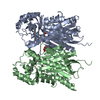
| ||||||||
|---|---|---|---|---|---|---|---|---|---|
| 1 |
| ||||||||
| Unit cell |
| ||||||||
| Noncrystallographic symmetry (NCS) | NCS oper: (Code: given Matrix: (-0.818, 0.4, 0.413), Vector: |
-Components
| #1: Protein | Mass: 44509.203 Da / Num. of mol.: 2 Source method: isolated from a genetically manipulated source Details: COVALENT LINK BETWEEN C 194 AND THE ACETYL GROUP OF THE FATTY ACID LIGAND Source: (gene. exp.) ECTOCARPUS SILICULOSUS (eukaryote) / Strain: EC32 / Production host:  References: UniProt: D8LJ35, Transferases; Acyltransferases; Transferring groups other than aminoacyl groups #2: Chemical |   Mass: 104.061 Da / Num. of mol.: 2 / Source method: obtained synthetically / Formula: C3H4O4 Mass: 104.061 Da / Num. of mol.: 2 / Source method: obtained synthetically / Formula: C3H4O4#3: Chemical |   Mass: 304.467 Da / Num. of mol.: 2 / Source method: obtained synthetically / Formula: C20H32O2 Mass: 304.467 Da / Num. of mol.: 2 / Source method: obtained synthetically / Formula: C20H32O2#4: Water | ChemComp-HOH / |  Mass: 18.015 Da / Num. of mol.: 343 / Source method: isolated from a natural source / Formula: H2O Mass: 18.015 Da / Num. of mol.: 343 / Source method: isolated from a natural source / Formula: H2OHas protein modification | Y | Nonpolymer details | ARACHIDONI | |
|---|
-Experimental details
-Experiment
| Experiment | Method: X-RAY DIFFRACTION / Number of used crystals: 1 |
|---|
- Sample preparation
| Crystal | Density Matthews: 2.39 Å3/Da / Density % sol: 48.5 % / Description: NONE |
|---|---|
| Crystal grow | Temperature: 292 K / Method: vapor diffusion, hanging drop / pH: 8 Details: 3 MICROLITER DROPS OF PROTEIN AT 7 MG/ML WERE MIXED WITH 1.5 MICROLITERS OF CRYSTALLIZATION BUFFER (0.1 M BICINE PH 8, 18% PEG 6000) USING THE HANGING DROP VAPOR DIFFUSION METHOD AT 19 DEGREES CELSIUS |
-Data collection
| Diffraction | Mean temperature: 100 K |
|---|---|
| Diffraction source | Source: SYNCHROTRON / Site: ESRF  / Beamline: ID23-1 / Wavelength: 0.9792 / Beamline: ID23-1 / Wavelength: 0.9792 |
| Detector | Type: ADSC QUANTUM Q4 / Detector: CCD / Date: Feb 23, 2011 / Details: MIRRORS |
| Radiation | Protocol: SINGLE WAVELENGTH / Monochromatic (M) / Laue (L): M / Scattering type: x-ray |
| Radiation wavelength | Wavelength: 0.9792 Å / Relative weight: 1 |
| Reflection | Resolution: 2.85→49 Å / Num. obs: 18963 / % possible obs: 91.7 % / Observed criterion σ(I): 0 / Redundancy: 4.2 % / Rmerge(I) obs: 0.1 / Net I/σ(I): 8.5 |
| Reflection shell | Resolution: 2.85→3.02 Å / Redundancy: 4.3 % / Rmerge(I) obs: 0.33 / Mean I/σ(I) obs: 3.4 / % possible all: 77.4 |
- Processing
| Software |
| ||||||||||||||||||||||||||||||||||||||||||||||||||||||||||||||||||||||||||||||||||||||||||||||||||||||||||||||||||||||||||||||||||||||||||||||||||||||||||||||||||||||||||||||||||||||
|---|---|---|---|---|---|---|---|---|---|---|---|---|---|---|---|---|---|---|---|---|---|---|---|---|---|---|---|---|---|---|---|---|---|---|---|---|---|---|---|---|---|---|---|---|---|---|---|---|---|---|---|---|---|---|---|---|---|---|---|---|---|---|---|---|---|---|---|---|---|---|---|---|---|---|---|---|---|---|---|---|---|---|---|---|---|---|---|---|---|---|---|---|---|---|---|---|---|---|---|---|---|---|---|---|---|---|---|---|---|---|---|---|---|---|---|---|---|---|---|---|---|---|---|---|---|---|---|---|---|---|---|---|---|---|---|---|---|---|---|---|---|---|---|---|---|---|---|---|---|---|---|---|---|---|---|---|---|---|---|---|---|---|---|---|---|---|---|---|---|---|---|---|---|---|---|---|---|---|---|---|---|---|---|
| Refinement | Method to determine structure: MOLECULAR REPLACEMENT Starting model: PDB ENTRY 1TED Resolution: 2.85→49.4 Å / Cor.coef. Fo:Fc: 0.915 / Cor.coef. Fo:Fc free: 0.897 / SU B: 24.507 / SU ML: 0.276 / Cross valid method: THROUGHOUT / ESU R Free: 0.406 / Stereochemistry target values: MAXIMUM LIKELIHOOD Details: HYDROGENS HAVE BEEN ADDED IN THE RIDING POSITIONS. HYDROGENS HAVE BEEN USED IF PRESENT IN THE INPUT. DISORDERED RESIDUES AND LIGAND WERE MODELED WITH OCCUPANCY LOWER THAN 1.00.
| ||||||||||||||||||||||||||||||||||||||||||||||||||||||||||||||||||||||||||||||||||||||||||||||||||||||||||||||||||||||||||||||||||||||||||||||||||||||||||||||||||||||||||||||||||||||
| Solvent computation | Ion probe radii: 0.8 Å / Shrinkage radii: 0.8 Å / VDW probe radii: 1.2 Å / Solvent model: MASK | ||||||||||||||||||||||||||||||||||||||||||||||||||||||||||||||||||||||||||||||||||||||||||||||||||||||||||||||||||||||||||||||||||||||||||||||||||||||||||||||||||||||||||||||||||||||
| Displacement parameters | Biso mean: 39.972 Å2
| ||||||||||||||||||||||||||||||||||||||||||||||||||||||||||||||||||||||||||||||||||||||||||||||||||||||||||||||||||||||||||||||||||||||||||||||||||||||||||||||||||||||||||||||||||||||
| Refinement step | Cycle: LAST / Resolution: 2.85→49.4 Å
| ||||||||||||||||||||||||||||||||||||||||||||||||||||||||||||||||||||||||||||||||||||||||||||||||||||||||||||||||||||||||||||||||||||||||||||||||||||||||||||||||||||||||||||||||||||||
| Refine LS restraints |
|