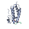
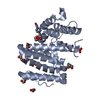
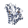
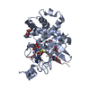
- PDB-4au7: The structure of the Suv4-20h2 ternary complex with histone H4 -
+
Open data
ID or keywords:
Loading...
-
Basic information
Entry
Database: PDB / ID: 4au7
Title
The structure of the Suv4-20h2 ternary complex with histone H4
Components
HISTONE H4 PEPTIDE
HISTONE-LYSINE N-METHYLTRANSFERASE SUV420H2
Keywords
TRANSFERASE / EPIGENETICS
Function / homology
Function and homology information
Deposition of new CENPA-containing nucleosomes at the centromere / Inhibition of DNA recombination at telomere / SUMOylation of chromatin organization proteins / [histone H4]-N-methyl-L-lysine20 N-methyltransferase / DNA Damage/Telomere Stress Induced Senescence / G2/M DNA damage checkpoint / Regulation of PD-L1(CD274) transcription / Regulation of endogenous retroelements by KRAB-ZFP proteins / Condensation of Prophase Chromosomes / HDMs demethylate histones ...Deposition of new CENPA-containing nucleosomes at the centromere / Inhibition of DNA recombination at telomere / SUMOylation of chromatin organization proteins / [histone H4]-N-methyl-L-lysine20 N-methyltransferase / DNA Damage/Telomere Stress Induced Senescence / G2/M DNA damage checkpoint / Regulation of PD-L1(CD274) transcription / Regulation of endogenous retroelements by KRAB-ZFP proteins / Condensation of Prophase Chromosomes / HDMs demethylate histones / Nonhomologous End-Joining (NHEJ) / Negative Regulation of CDH1 Gene Transcription / Recognition and association of DNA glycosylase with site containing an affected purine / [histone H4]-lysine20 N-methyltransferase / histone H4K20 monomethyltransferase activity / Cleavage of the damaged purine / PRC2 methylates histones and DNA / HATs acetylate histones / histone H4K20me methyltransferase activity / histone H4K20 methyltransferase activity / MLL4 and MLL3 complexes regulate expression of PPARG target genes in adipogenesis and hepatic steatosis / PKMTs methylate histone lysines / Processing of DNA double-strand break ends / Recruitment and ATM-mediated phosphorylation of repair and signaling proteins at DNA double strand breaks / RUNX1 regulates genes involved in megakaryocyte differentiation and platelet function / RMTs methylate histone arginines / positive regulation of isotype switching / Estrogen-dependent gene expression / condensed chromosome, centromeric region / S-adenosyl-L-methionine binding / positive regulation of double-strand break repair via nonhomologous end joining / pericentric heterochromatin / protein localization to CENP-A containing chromatin / heterochromatin / CENP-A containing nucleosome / structural constituent of chromatin / nucleosome / nucleosome assembly / chromatin organization / methylation / histone binding / protein heterodimerization activity / DNA repair / chromatin binding / protein-containing complex / DNA binding / extracellular exosome / nucleoplasm / metal ion binding / nucleus Similarity search - Function
KMT5C , SET domain / Histone-lysine N-methyltransferase / Suv4-20 family, animal / Histone-lysine N-methyltransferase Suv4-20/Set9 / Histone-lysine N-methyltransferase, N-terminal domain / Histone-lysine N-methyltransferase (EC 2.1.1.43) family profile. / Beta-clip-like / SET domain / SET (Su(var)3-9, Enhancer-of-zeste, Trithorax) domain / SET domain ...KMT5C , SET domain / Histone-lysine N-methyltransferase / Suv4-20 family, animal / Histone-lysine N-methyltransferase Suv4-20/Set9 / Histone-lysine N-methyltransferase, N-terminal domain / Histone-lysine N-methyltransferase (EC 2.1.1.43) family profile. / Beta-clip-like / SET domain / SET (Su(var)3-9, Enhancer-of-zeste, Trithorax) domain / SET domain / SET domain profile. / SET domain superfamily / SET domain / Beta Complex / TATA box binding protein associated factor / TATA box binding protein associated factor (TAF), histone-like fold domain / Histone H4, conserved site / Histone H4 signature. / Histone H4 / Histone H4 / CENP-T/Histone H4, histone fold / Centromere kinetochore component CENP-T histone fold / Histone-fold / Arc Repressor Mutant, subunit A / Orthogonal Bundle / Mainly Beta / Mainly Alpha Similarity search - Domain/homology
Resolution: 2.07→55.33 Å / Cor.coef. Fo:Fc: 0.952 / Cor.coef. Fo:Fc free: 0.925 / SU B: 8.589 / SU ML: 0.123 / Cross valid method: THROUGHOUT / ESU R: 0.217 / ESU R Free: 0.186 / Stereochemistry target values: MAXIMUM LIKELIHOOD Details: HYDROGENS HAVE BEEN ADDED IN THE RIDING POSITIONS. THE C-TERMINUS OF MOECULE B IS DISORDERED
Rfactor
Num. reflection
% reflection
Selection details
Rfree
0.24039
1570
5 %
RANDOM
Rwork
0.19255
-
-
-
obs
0.19498
29593
97.05 %
-
Solvent computation
Ion probe radii: 0.8 Å / Shrinkage radii: 0.8 Å / VDW probe radii: 1.2 Å / Solvent model: MASK
 Movie
Movie Controller


 Open data
 Basic information
 Components
 Keywords
 Function and homology information
 X-RAY DIFFRACTION /
  Authors
 Citation
 Structure visualization
 Downloads & links
 Other downloads
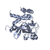


 PDBj
PDBj


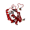
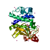
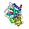
 Assembly


 Mass: 384.411 Da / Num. of mol.: 1 / Source method: obtained synthetically / Formula: C14H20N6O5S
 Mass: 65.409 Da / Num. of mol.: 3 / Source method: obtained synthetically / Formula: Zn
 Mass: 62.068 Da / Num. of mol.: 16 / Source method: obtained synthetically / Formula: C2H6O2
 Sample preparation
 / Beamline: I02 / Wavelength: 1.0081
  Processing