 Movie
Movie Controller

[English] 日本語
 Yorodumi
- PDB-1urh: The "Rhodanese" fold and catalytic mechanism of 3-mercaptopyruvat... -
+ Open data

- Basic information
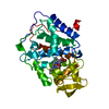
| Entry | Database: PDB / ID: 1urh | ||||||
|---|---|---|---|---|---|---|---|
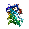
| Title | The "Rhodanese" fold and catalytic mechanism of 3-mercaptopyruvate sulfotransferases: Crystal structure of SseA from Escherichia coli | ||||||
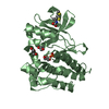
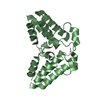
 Components Components | 3-MERCAPTOPYRUVATE SULFURTRANSFERASE | ||||||
 Keywords Keywords | TRANSFERASE / SULFUR-TRANSFERASE / RHODANESE | ||||||
| Function / homology |  Function and homology information Function and homology information3-mercaptopyruvate sulfurtransferase / 3-mercaptopyruvate sulfurtransferase activity / thiosulfate-cyanide sulfurtransferase activity / DNA protection / response to oxidative stress / response to antibiotic / cytosol Similarity search - Function | ||||||
| Biological species |  | ||||||
| Method |  X-RAY DIFFRACTION / SYNCHROTRON / MOLECULAR REPLACEMENT / Resolution: 2.8 Å X-RAY DIFFRACTION / SYNCHROTRON / MOLECULAR REPLACEMENT / Resolution: 2.8 Å | ||||||
 Authors Authors | Spallarossa, A. / Forlani, F. / Carpen, A. / Armirotti, A. / Pagani, S. / Bolognesi, M. / Bordo, D. | ||||||
 Citation Citation | Journal: J.Mol.Biol. / Year: 2004 Title: The "Rhodanese" Fold and Catalytic Mechanism of 3-Mercaptopyruvate Sulfurtransferases: Crystal Structure of Ssea from Escherichia Coli Authors: Spallarossa, A. / Forlani, F. / Carpen, A. / Armirotti, A. / Pagani, S. / Bolognesi, M. / Bordo, D. | ||||||
| History |
|
- Structure visualization
| Structure viewer | Molecule: MolmilJmol/JSmol |
|---|
- Downloads & links
-Download
| PDBx/mmCIF format | 1urh.cif.gz | 111.1 KB | Display | PDBx/mmCIF format |
|---|---|---|---|---|
| PDB format | pdb1urh.ent.gz | 86.9 KB | Display | PDB format |
| PDBx/mmJSON format | 1urh.json.gz | Tree view | PDBx/mmJSON format | |
| Others |  Other downloads Other downloads |
-Validation report
| Arichive directory | https://data.pdbj.org/pub/pdb/validation_reports/ur/1urhftp://data.pdbj.org/pub/pdb/validation_reports/ur/1urh | HTTPS FTP |
|---|
-Related structure data
| Related structure data |  1rhsS S: Starting model for refinement |
|---|---|
| Similar structure data |


-Links
 PDBj
PDBj
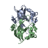
- Assembly
| Deposited unit | 
| ||||||||
|---|---|---|---|---|---|---|---|---|---|
| 1 | 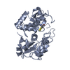
| ||||||||
| 2 | 
| ||||||||
| Unit cell |
|
-Components
| #1: Protein | Mass: 30741.596 Da / Num. of mol.: 2 Source method: isolated from a genetically manipulated source Details: RESIDUES A186-A189 AND B181-B191 ARE NOT INCLUDED IN THE MODEL. Source: (gene. exp.) References: UniProt: P31142, 3-mercaptopyruvate sulfurtransferase #2: Chemical | ChemComp-SO3 / | 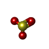  Mass: 80.063 Da / Num. of mol.: 1 / Source method: obtained synthetically / Formula: SO3 Mass: 80.063 Da / Num. of mol.: 1 / Source method: obtained synthetically / Formula: SO3#3: Water | ChemComp-HOH / |  Mass: 18.015 Da / Num. of mol.: 68 / Source method: isolated from a natural source / Formula: H2O Mass: 18.015 Da / Num. of mol.: 68 / Source method: isolated from a natural source / Formula: H2OCompound details | 3-MERCAPTOPY | Has protein modification | Y | |
|---|
-Experimental details
-Experiment
| Experiment | Method: X-RAY DIFFRACTION / Number of used crystals: 1 |
|---|
- Sample preparation
| Crystal | Density Matthews: 3.5 Å3/Da / Density % sol: 65 % | ||||||||||||||||||||||||||||||||||||||||||||||||||||||||
|---|---|---|---|---|---|---|---|---|---|---|---|---|---|---|---|---|---|---|---|---|---|---|---|---|---|---|---|---|---|---|---|---|---|---|---|---|---|---|---|---|---|---|---|---|---|---|---|---|---|---|---|---|---|---|---|---|---|
| Crystal grow | pH: 6.5 / Details: pH 6.50 | ||||||||||||||||||||||||||||||||||||||||||||||||||||||||
| Crystal grow | *PLUS Temperature: 294 K / pH: 8 / Method: vapor diffusion, hanging drop / Details: Spallarossa, A., (2003) Acta Cryst., D59, 168. | ||||||||||||||||||||||||||||||||||||||||||||||||||||||||
| Components of the solutions | *PLUS
|
-Data collection
| Diffraction | Mean temperature: 100 K |
|---|---|
| Diffraction source | Source: SYNCHROTRON / Site: ESRF  / Beamline: ID14-4 / Wavelength: 0.9393 / Beamline: ID14-4 / Wavelength: 0.9393 |
| Detector | Date: Jul 15, 2002 |
| Radiation | Protocol: SINGLE WAVELENGTH / Monochromatic (M) / Laue (L): M / Scattering type: x-ray |
| Radiation wavelength | Wavelength: 0.9393 Å / Relative weight: 1 |
| Reflection | Resolution: 2.8→40 Å / Num. obs: 21524 / % possible obs: 96 % / Observed criterion σ(I): 2 / Redundancy: 14 % / Rmerge(I) obs: 0.066 / Net I/σ(I): 16 |
| Reflection shell | Resolution: 2.8→2.85 Å / Rmerge(I) obs: 0.483 / Mean I/σ(I) obs: 2 / % possible all: 97.9 |
| Reflection | *PLUS Highest resolution: 2.8 Å / Lowest resolution: 40 Å / % possible obs: 95.6 % / Num. measured all: 312079 / Rmerge(I) obs: 0.066 |
| Reflection shell | *PLUS % possible obs: 97.9 % / Rmerge(I) obs: 0.483 / Mean I/σ(I) obs: 2.2 |
- Processing
| Software |
| ||||||||||||||||||||||||||||||||||||||||||||||||||||||||||||
|---|---|---|---|---|---|---|---|---|---|---|---|---|---|---|---|---|---|---|---|---|---|---|---|---|---|---|---|---|---|---|---|---|---|---|---|---|---|---|---|---|---|---|---|---|---|---|---|---|---|---|---|---|---|---|---|---|---|---|---|---|---|
| Refinement | Method to determine structure: MOLECULAR REPLACEMENT Starting model: PDB ENTRY 1RHS Resolution: 2.8→40 Å / Cross valid method: THROUGHOUT / σ(F): 2
| ||||||||||||||||||||||||||||||||||||||||||||||||||||||||||||
| Refinement step | Cycle: LAST / Resolution: 2.8→40 Å
| ||||||||||||||||||||||||||||||||||||||||||||||||||||||||||||
| Refine LS restraints |
| ||||||||||||||||||||||||||||||||||||||||||||||||||||||||||||
| Refinement | *PLUS % reflection Rfree: 5 % | ||||||||||||||||||||||||||||||||||||||||||||||||||||||||||||
| Solvent computation | *PLUS | ||||||||||||||||||||||||||||||||||||||||||||||||||||||||||||
| Displacement parameters | *PLUS | ||||||||||||||||||||||||||||||||||||||||||||||||||||||||||||
| LS refinement shell | *PLUS Highest resolution: 2.8 Å / Lowest resolution: 2.9 Å / Rfactor Rfree: 0.447 / Rfactor Rwork: 0.385 |
