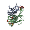
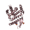
[English] 日本語
Yorodumi
- PDB-4aoc: crystal structure of BC2L-A Lectin from Burkolderia cenocepacia i... -

+
Open data


ID or keywords:

Loading...

-
Basic information

Entry
Database: PDB / ID: 4aoc
Titlecrystal structure of BC2L-A Lectin from Burkolderia cenocepacia in complex with methyl-heptoside
ComponentsBC2L-A LECTIN
KeywordsHYDROLASE
Function / homology
Function and homology information


Calcium-mediated lectin / Calcium-mediated lectin / Calcium-mediated lectin superfamily / Fucose-binding lectin II (PA-IIL) / Jelly Rolls / Sandwich / Mainly Beta
Similarity search - Domain/homology
methyl L-glycero-alpha-D-manno-heptopyranoside / PA-IIL domain-containing protein / PA-IIL domain-containing protein
Similarity search - Component
Biological speciesBURKHOLDERIA CENOCEPACIA (bacteria)
MethodX-RAY DIFFRACTION / SYNCHROTRON / MOLECULAR REPLACEMENT / Resolution: 2.7 Å
AuthorsMarchetti, R. / Malinovska, L. / Lameignere, E. / deCastro, C. / Cioci, G. / Kosma, P. / Wimmerova, M. / Molinaro, A. / Imberty, A. / Silipo, A.
CitationJournal: Glycobiology / Year: 2012
Title: Burkholderia Cenocepacia Lectin a Binding to Heptoses from the Bacterial Lipopolysaccharide.
Authors: Marchetti, R. / Malinovska, L. / Lameignere, E. / Adamova, L. / De Castro, C. / Cioci, G. / Stanetty, C. / Kosma, P. / Molinaro, A. / Wimmerova, M. / Imberty, A. / Silipo, A.
History
DepositionMar 26, 2012Deposition site: PDBE / Processing site: PDBE
Revision 1.0Aug 15, 2012Provider: repository / Type: Initial release
Revision 1.1Oct 3, 2012Group: Database references
Revision 1.2Nov 19, 2014Group: Data collection
Revision 1.3Jul 29, 2020Group: Data collection / Derived calculations / Other
Category: chem_comp / pdbx_database_status ...chem_comp / pdbx_database_status / pdbx_struct_conn_angle / struct_conn / struct_site / struct_site_gen
Item: _chem_comp.mon_nstd_flag / _pdbx_database_status.status_code_sf ..._chem_comp.mon_nstd_flag / _pdbx_database_status.status_code_sf / _pdbx_struct_conn_angle.ptnr1_auth_asym_id / _pdbx_struct_conn_angle.ptnr1_auth_comp_id / _pdbx_struct_conn_angle.ptnr1_auth_seq_id / _pdbx_struct_conn_angle.ptnr1_label_asym_id / _pdbx_struct_conn_angle.ptnr1_label_atom_id / _pdbx_struct_conn_angle.ptnr1_label_comp_id / _pdbx_struct_conn_angle.ptnr1_label_seq_id / _pdbx_struct_conn_angle.ptnr1_symmetry / _pdbx_struct_conn_angle.ptnr2_auth_asym_id / _pdbx_struct_conn_angle.ptnr2_auth_seq_id / _pdbx_struct_conn_angle.ptnr2_label_asym_id / _pdbx_struct_conn_angle.ptnr3_auth_asym_id / _pdbx_struct_conn_angle.ptnr3_auth_comp_id / _pdbx_struct_conn_angle.ptnr3_auth_seq_id / _pdbx_struct_conn_angle.ptnr3_label_asym_id / _pdbx_struct_conn_angle.ptnr3_label_atom_id / _pdbx_struct_conn_angle.ptnr3_label_comp_id / _pdbx_struct_conn_angle.ptnr3_label_seq_id / _pdbx_struct_conn_angle.ptnr3_symmetry / _pdbx_struct_conn_angle.value / _struct_conn.pdbx_dist_value / _struct_conn.ptnr1_auth_asym_id / _struct_conn.ptnr1_auth_comp_id / _struct_conn.ptnr1_auth_seq_id / _struct_conn.ptnr1_label_asym_id / _struct_conn.ptnr1_label_atom_id / _struct_conn.ptnr1_label_comp_id / _struct_conn.ptnr1_label_seq_id / _struct_conn.ptnr1_symmetry / _struct_conn.ptnr2_auth_asym_id / _struct_conn.ptnr2_auth_comp_id / _struct_conn.ptnr2_auth_seq_id / _struct_conn.ptnr2_label_asym_id / _struct_conn.ptnr2_label_atom_id / _struct_conn.ptnr2_label_comp_id / _struct_conn.ptnr2_label_seq_id / _struct_conn.ptnr2_symmetry
Description: Carbohydrate remediation / Provider: repository / Type: Remediation
Revision 1.4Dec 20, 2023Group: Data collection / Database references ...Data collection / Database references / Refinement description / Structure summary
Category: chem_comp / chem_comp_atom ...chem_comp / chem_comp_atom / chem_comp_bond / database_2 / pdbx_initial_refinement_model
Item: _chem_comp.pdbx_synonyms / _database_2.pdbx_DOI / _database_2.pdbx_database_accession

-
Structure visualization

Structure viewerMolecule:
MolmilJmol/JSmol

Downloads & links

-
Assembly

Deposited unit
A: BC2L-A LECTIN
B: BC2L-A LECTIN
C: BC2L-A LECTIN
D: BC2L-A LECTIN
E: BC2L-A LECTIN
hetero molecules


Theoretical massNumber of molelcules
Total (without water)72,13031
Polymers69,5515
Non-polymers2,57926
Water2,018112
1
A: BC2L-A LECTIN
B: BC2L-A LECTIN
hetero molecules


Theoretical massNumber of molelcules
Total (without water)28,71711
Polymers27,8202
Non-polymers8979
Water362
TypeNameSymmetry operationNumber
identity operation1_555x,y,z1
Buried area3640 Å2
ΔGint-73.4 kcal/mol
Surface area10760 Å2
MethodPISA
2
C: BC2L-A LECTIN
E: BC2L-A LECTIN
hetero molecules


Theoretical massNumber of molelcules
Total (without water)28,81312
Polymers27,8202
Non-polymers99310
Water362
TypeNameSymmetry operationNumber
identity operation1_555x,y,z1
Buried area4050 Å2
ΔGint-103.9 kcal/mol
Surface area10880 Å2
MethodPISA
3
D: BC2L-A LECTIN
hetero molecules

D: BC2L-A LECTIN
hetero molecules


Theoretical massNumber of molelcules
Total (without water)29,19816
Polymers27,8202
Non-polymers1,37714
Water362
TypeNameSymmetry operationNumber
identity operation1_555x,y,z1
crystal symmetry operation4_555x,-y,-z1
Buried area3700 Å2
ΔGint-83.1 kcal/mol
Surface area10520 Å2
MethodPISA
Unit cell
Length a, b, c (Å)50.000, 185.140, 186.210
Angle α, β, γ (deg.)90.00, 90.00, 90.00
Int Tables number20
Space group name H-MC2221
Components on special symmetry positions
IDModelComponents
11D-1133-

SO4

-
Components

#1: Protein
BC2L-A LECTIN


Mass: 13910.230 Da / Num. of mol.: 5
Source method: isolated from a genetically manipulated source
Source: (gene. exp.) BURKHOLDERIA CENOCEPACIA (bacteria) / Strain: AU 1054 / Production host: ESCHERICHIA COLI (E. coli) / References: UniProt: Q1BKJ8, UniProt: A0A0H2Y045*PLUS
#2: Chemical
ChemComp-CA / CALCIUM ION


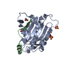
Mass: 40.078 Da / Num. of mol.: 10 / Source method: obtained synthetically / Formula: Ca
#3: Sugar
ChemComp-A1Q / methyl L-glycero-alpha-D-manno-heptopyranoside / ALPHA-METHYL HEPTOPYRANOSE / methyl L-glycero-alpha-D-manno-heptoside / methyl L-glycero-D-manno-heptoside / methyl L-glycero-manno-heptoside


Type: D-saccharide / Mass: 224.208 Da / Num. of mol.: 5
Source method: isolated from a genetically manipulated source
Formula: C8H16O7
#4: Chemical
ChemComp-SO4 / SULFATE ION


Mass: 96.063 Da / Num. of mol.: 11 / Source method: obtained synthetically / Formula: SO4
#5: Water ChemComp-HOH / water


Mass: 18.015 Da / Num. of mol.: 112 / Source method: isolated from a natural source / Formula: H2O

-
Experimental details

-
Experiment

ExperimentMethod: X-RAY DIFFRACTION / Number of used crystals: 1

-
Sample preparation

CrystalDensity Matthews: 2.95 Å3/Da / Density % sol: 58.3 % / Description: NONE

-
Data collection

DiffractionMean temperature: 100 K
Diffraction sourceSource: SYNCHROTRON / Site: ESRF / Beamline: ID23-2 / Wavelength: 0.8726
DetectorType: MARMOSAIC 225 mm CCD / Detector: CCD / Details: KB MIRROR
RadiationMonochromator: SI111 / Protocol: SINGLE WAVELENGTH / Monochromatic (M) / Laue (L): M / Scattering type: x-ray
Radiation wavelengthWavelength: 0.8726 Å / Relative weight: 1
ReflectionResolution: 2.7→48.27 Å / Num. obs: 24256 / % possible obs: 99.6 % / Observed criterion σ(I): 2 / Redundancy: 4.8 % / Rmerge(I) obs: 0.14 / Net I/σ(I): 10.2
Reflection shellResolution: 2.7→2.85 Å / Redundancy: 4.6 % / Rmerge(I) obs: 0.52 / Mean I/σ(I) obs: 2.9 / % possible all: 81.7

-
Processing

Software
NameVersionClassification
REFMAC5.6.0117refinement
XDSdata reduction
SCALAdata scaling
MOLREPphasing
RefinementMethod to determine structure: MOLECULAR REPLACEMENT
Starting model: PDB ENTRY 2WR9
Resolution: 2.7→93.11 Å / Cor.coef. Fo:Fc: 0.928 / Cor.coef. Fo:Fc free: 0.88 / SU B: 10.79 / SU ML: 0.218 / Cross valid method: THROUGHOUT / ESU R: 0.605 / ESU R Free: 0.319 / Stereochemistry target values: MAXIMUM LIKELIHOOD
Details: HYDROGENS HAVE BEEN ADDED IN THE RIDING POSITIONS. U VALUES REFINED INDIVIDUALLY
RfactorNum. reflection% reflectionSelection details
Rfree0.2532 1236 5.1 %RANDOM
Rwork0.19228 ---
obs0.19545 23002 99.49 %-
Solvent computationIon probe radii: 0.8 Å / Shrinkage radii: 0.8 Å / VDW probe radii: 1.2 Å / Solvent model: MASK
Displacement parametersBiso mean: 20.226 Å2
Baniso -1Baniso -2Baniso -3
1-0.8 Å20 Å20 Å2
2--0 Å20 Å2
3----0.81 Å2
Refinement stepCycle: LAST / Resolution: 2.7→93.11 Å
ProteinNucleic acidLigandSolventTotal
Num. atoms4543 0 140 112 4795
Refine LS restraints
Refine-IDTypeDev idealDev ideal targetNumber
X-RAY DIFFRACTIONr_bond_refined_d0.0110.024813
X-RAY DIFFRACTIONr_bond_other_d0.0010.023215
X-RAY DIFFRACTIONr_angle_refined_deg1.5691.9656553
X-RAY DIFFRACTIONr_angle_other_deg0.90137843
X-RAY DIFFRACTIONr_dihedral_angle_1_deg7.8455601
X-RAY DIFFRACTIONr_dihedral_angle_2_deg37.50224.612232
X-RAY DIFFRACTIONr_dihedral_angle_3_deg17.08615709
X-RAY DIFFRACTIONr_dihedral_angle_4_deg17.1441526
X-RAY DIFFRACTIONr_chiral_restr0.0870.2718
X-RAY DIFFRACTIONr_gen_planes_refined0.0060.0215412
X-RAY DIFFRACTIONr_gen_planes_other0.0010.02966
X-RAY DIFFRACTIONr_nbd_refined
X-RAY DIFFRACTIONr_nbd_other
X-RAY DIFFRACTIONr_nbtor_refined
X-RAY DIFFRACTIONr_nbtor_other
X-RAY DIFFRACTIONr_xyhbond_nbd_refined
X-RAY DIFFRACTIONr_xyhbond_nbd_other
X-RAY DIFFRACTIONr_metal_ion_refined
X-RAY DIFFRACTIONr_metal_ion_other
X-RAY DIFFRACTIONr_symmetry_vdw_refined
X-RAY DIFFRACTIONr_symmetry_vdw_other
X-RAY DIFFRACTIONr_symmetry_hbond_refined
X-RAY DIFFRACTIONr_symmetry_hbond_other
X-RAY DIFFRACTIONr_symmetry_metal_ion_refined
X-RAY DIFFRACTIONr_symmetry_metal_ion_other
X-RAY DIFFRACTIONr_mcbond_it
X-RAY DIFFRACTIONr_mcbond_other
X-RAY DIFFRACTIONr_mcangle_it
X-RAY DIFFRACTIONr_mcangle_other
X-RAY DIFFRACTIONr_scbond_it
X-RAY DIFFRACTIONr_scbond_other
X-RAY DIFFRACTIONr_scangle_it
X-RAY DIFFRACTIONr_scangle_other
X-RAY DIFFRACTIONr_long_range_B_refined
X-RAY DIFFRACTIONr_long_range_B_other
X-RAY DIFFRACTIONr_rigid_bond_restr
X-RAY DIFFRACTIONr_sphericity_free
X-RAY DIFFRACTIONr_sphericity_bonded
LS refinement shellResolution: 2.7→2.77 Å / Total num. of bins used: 20
RfactorNum. reflection% reflection
Rfree0.452 78 -
Rwork0.307 1514 -
obs--95.85 %

+
About Yorodumi

-
News

-
Feb 9, 2022. New format data for meta-information of EMDB entries

New format data for meta-information of EMDB entries

  • Version 3 of the EMDB header file is now the official format.
  • The previous official version 1.9 will be removed from the archive.

Related info.:EMDB header

External links:wwPDB to switch to version 3 of the EMDB data model

-
Aug 12, 2020. Covid-19 info

Covid-19 info

URL: https://pdbjlvh1.pdbj.org/emnavi/covid19.php

New page: Covid-19 featured information page in EM Navigator.

Related info.:Covid-19 info / Mar 5, 2020. Novel coronavirus structure data

+
Mar 5, 2020. Novel coronavirus structure data

Novel coronavirus structure data

Related info.:Yorodumi Speices / Aug 12, 2020. Covid-19 info

External links:COVID-19 featured content - PDBj / Molecule of the Month (242):Coronavirus Proteases

+
Jan 31, 2019. EMDB accession codes are about to change! (news from PDBe EMDB page)

EMDB accession codes are about to change! (news from PDBe EMDB page)

  • The allocation of 4 digits for EMDB accession codes will soon come to an end. Whilst these codes will remain in use, new EMDB accession codes will include an additional digit and will expand incrementally as the available range of codes is exhausted. The current 4-digit format prefixed with “EMD-” (i.e. EMD-XXXX) will advance to a 5-digit format (i.e. EMD-XXXXX), and so on. It is currently estimated that the 4-digit codes will be depleted around Spring 2019, at which point the 5-digit format will come into force.
  • The EM Navigator/Yorodumi systems omit the EMD- prefix.

Related info.:Q: What is EMD? / ID/Accession-code notation in Yorodumi/EM Navigator

External links:EMDB Accession Codes are Changing Soon! / Contact to PDBj

+
Jul 12, 2017. Major update of PDB

Major update of PDB

  • wwPDB released updated PDB data conforming to the new PDBx/mmCIF dictionary.
  • This is a major update changing the version number from 4 to 5, and with Remediation, in which all the entries are updated.
  • In this update, many items about electron microscopy experimental information are reorganized (e.g. em_software).
  • Now, EM Navigator and Yorodumi are based on the updated data.

External links:wwPDB Remediation / Enriched Model Files Conforming to OneDep Data Standards Now Available in the PDB FTP Archive

-
Yorodumi

Thousand views of thousand structures

  • Yorodumi is a browser for structure data from EMDB, PDB, SASBDB, etc.
  • This page is also the successor to EM Navigator detail page, and also detail information page/front-end page for Omokage search.
  • The word "yorodu" (or yorozu) is an old Japanese word meaning "ten thousand". "mi" (miru) is to see.

Related info.:EMDB / PDB / SASBDB / Comparison of 3 databanks / Yorodumi Search / Aug 31, 2016. New EM Navigator & Yorodumi / Yorodumi Papers / Jmol/JSmol / Function and homology information / Changes in new EM Navigator and Yorodumi

Read more