 Movie
Movie Controller


+ Open data

- Basic information
| Entry | Database: PDB / ID: 3c1u | ||||||
|---|---|---|---|---|---|---|---|
| Title | D192N mutant of Rhamnogalacturonan acetylesterase | ||||||
 Components Components | Rhamnogalacturonan acetylesterase | ||||||
 Keywords Keywords | HYDROLASE / SGNH hydrolase / pectin degradation / Glycoprotein | ||||||
| Function / homology |  Function and homology information Function and homology informationrhamnogalacturonan acetylesterase / hydrolase activity, acting on ester bonds Similarity search - Function | ||||||
| Biological species |  | ||||||
| Method |  X-RAY DIFFRACTION / SYNCHROTRON / MOLECULAR REPLACEMENT / Resolution: 1.33 Å X-RAY DIFFRACTION / SYNCHROTRON / MOLECULAR REPLACEMENT / Resolution: 1.33 Å | ||||||
 Authors Authors | Langkilde, A. / Lo Leggio, L. / Navarro Poulsen, J.C. / Molgaard, A. / Larsen, S. | ||||||
 Citation Citation | Journal: ACTA CRYSTALLOGR.,SECT.D / Year: 2008 Title: Short strong hydrogen bonds in proteins: a case study of rhamnogalacturonan acetylesterase Authors: Langkilde, A. / Kristensen, S.M. / Lo Leggio, L. / Jensen, J.H. / Houk, A.R. / Navarro Poulsen, J.C. / Kauppinen, S. / Larsen, S. #1: Journal: ACTA CRYSTALLOGR.,SECT.D / Year: 2004Title: Crystal packing in two pH-dependent crystal forms of rhamnogalacturonan acetylesterase Authors: Molgaard, A. / Larsen, S. #2: Journal: ACTA CRYSTALLOGR.,SECT.D / Year: 2002Title: A branched N-linked glycan at atomic resolution in the 1.12 A structure of rhamnogalacturonan acetylesterase Authors: Molgaard, A. / Larsen, S. #3: Journal: Structure / Year: 2000Title: Rhamnogalacturonan acetylesterase elucidates the structure and function of a new family of hydrolases Authors: Molgaard, A. / Kauppinen, S. / Larsen, S. #4: Journal: ACTA CRYSTALLOGR.,SECT.D / Year: 1998 Title: Crystallization and preliminary X-ray diffraction studies of the heterogeneously glycosylated enzyme rhamnogalacturonan acetylesterase from Aspergillus aculeatus Authors: Molgaard, A. / Petersen, J.F.W. / Kauppinen, S. / Dalboge, H. / Johnsen, A.H. / Navarro Poulsen, J.C. / Larsen, S. | ||||||
| History |
|
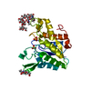
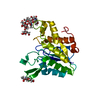
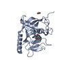
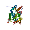
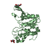
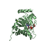
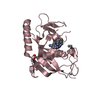
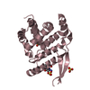
- Structure visualization
| Structure viewer | Molecule: MolmilJmol/JSmol |
|---|
- Downloads & links
-Download
| PDBx/mmCIF format | 3c1u.cif.gz | 118.4 KB | Display | PDBx/mmCIF format |
|---|---|---|---|---|
| PDB format | pdb3c1u.ent.gz | 92.2 KB | Display | PDB format |
| PDBx/mmJSON format | 3c1u.json.gz | Tree view | PDBx/mmJSON format | |
| Others |  Other downloads Other downloads |
-Validation report
| Arichive directory | https://data.pdbj.org/pub/pdb/validation_reports/c1/3c1uftp://data.pdbj.org/pub/pdb/validation_reports/c1/3c1u | HTTPS FTP |
|---|
-Related structure data
| Related structure data | 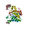 1k7cS S: Starting model for refinement |
|---|---|
| Similar structure data |


-Links
 PDBj
PDBj- Assembly
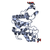
| Deposited unit | 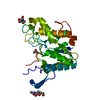
| ||||||||
|---|---|---|---|---|---|---|---|---|---|
| 1 |
| ||||||||
| Unit cell |
|
-Components
| #1: Protein | Mass: 24621.895 Da / Num. of mol.: 1 / Mutation: D192N Source method: isolated from a genetically manipulated source Source: (gene. exp.) References: UniProt: Q00017, Hydrolases; Acting on ester bonds; Carboxylic-ester hydrolases | ||||||
|---|---|---|---|---|---|---|---|
| #2: Sugar |   Type: D-saccharide, beta linking / Mass: 221.208 Da / Num. of mol.: 2 Type: D-saccharide, beta linking / Mass: 221.208 Da / Num. of mol.: 2Source method: isolated from a genetically manipulated source Formula: C8H15NO6 #3: Chemical | ChemComp-ACT / |   Mass: 59.044 Da / Num. of mol.: 1 / Source method: obtained synthetically / Formula: C2H3O2 Mass: 59.044 Da / Num. of mol.: 1 / Source method: obtained synthetically / Formula: C2H3O2#4: Water | ChemComp-HOH / |  Mass: 18.015 Da / Num. of mol.: 355 / Source method: isolated from a natural source / Formula: H2O Mass: 18.015 Da / Num. of mol.: 355 / Source method: isolated from a natural source / Formula: H2OHas protein modification | Y | |
-Experimental details
-Experiment
| Experiment | Method: X-RAY DIFFRACTION / Number of used crystals: 1 |
|---|
- Sample preparation
| Crystal | Density Matthews: 2.45 Å3/Da / Density % sol: 49.69 % |
|---|---|
| Crystal grow | Temperature: 298 K / Method: vapor diffusion, hanging drop / pH: 3 Details: PEG1500, sodium acetate, pH3.0, vapor diffusion, hanging drop, temperature 298K, VAPOR DIFFUSION, HANGING DROP |
-Data collection
| Diffraction | Mean temperature: 100 K |
|---|---|
| Diffraction source | Source: SYNCHROTRON / Site: ELETTRA  / Beamline: 5.2R / Wavelength: 1 Å / Beamline: 5.2R / Wavelength: 1 Å |
| Detector | Type: MARRESEARCH / Detector: CCD / Date: May 25, 2001 |
| Radiation | Protocol: SINGLE WAVELENGTH / Monochromatic (M) / Laue (L): M / Scattering type: x-ray |
| Radiation wavelength | Wavelength: 1 Å / Relative weight: 1 |
| Reflection | Resolution: 1.33→20 Å / Num. all: 55818 / Num. obs: 55818 / % possible obs: 99.5 % / Redundancy: 8.9 % / Rmerge(I) obs: 0.032 / Net I/σ(I): 31.8 |
| Reflection shell | Resolution: 1.33→1.35 Å / Redundancy: 2.3 % / Rmerge(I) obs: 0.198 / % possible all: 98.3 |
- Processing
| Software |
| |||||||||||||||||||||||||||||||||
|---|---|---|---|---|---|---|---|---|---|---|---|---|---|---|---|---|---|---|---|---|---|---|---|---|---|---|---|---|---|---|---|---|---|---|
| Refinement | Method to determine structure: MOLECULAR REPLACEMENT Starting model: PDB ENTRY 1K7C Resolution: 1.33→20 Å / Num. parameters: 19411 / Num. restraintsaints: 23615 / Cross valid method: FREE R / σ(F): 0 / Stereochemistry target values: Engh & Huber Details: One loop was very poorly defined in the electron density maps and as a consequence SER78 and THR81 are only modelled with an occupancy of 0.5. LEU79 and SER80 could not be positioned in the ...Details: One loop was very poorly defined in the electron density maps and as a consequence SER78 and THR81 are only modelled with an occupancy of 0.5. LEU79 and SER80 could not be positioned in the maps and are therefore not included in the model. The geometry and position of this loop is inflicted with higher uncertainty.
| |||||||||||||||||||||||||||||||||
| Solvent computation | Solvent model: MOEWS & KRETSINGER, J.MOL.BIOL.91(1973)201-228 | |||||||||||||||||||||||||||||||||
| Displacement parameters | Biso mean: 18.464 Å2 | |||||||||||||||||||||||||||||||||
| Refinement step | Cycle: LAST / Resolution: 1.33→20 Å
| |||||||||||||||||||||||||||||||||
| Refine LS restraints |
|
