 Movie
Movie Controller

[English] 日本語
 Yorodumi
- PDB-3nov: Crystal Structure of D17E Isocyanide Hydratase from Pseudomonas f... -
+ Open data

- Basic information
| Entry | Database: PDB / ID: 3nov | ||||||
|---|---|---|---|---|---|---|---|
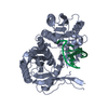
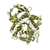
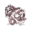
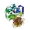
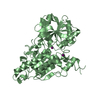
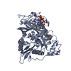
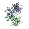
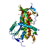
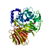
| Title | Crystal Structure of D17E Isocyanide Hydratase from Pseudomonas fluorescens | ||||||
 Components Components | ThiJ/PfpI family protein | ||||||
 Keywords Keywords | LYASE / DJ-1 superfamily / isocyanide hydratase / isonitrile hydratase | ||||||
| Function / homology |  Function and homology information Function and homology information | ||||||
| Biological species |  Pseudomonas fluorescens (bacteria) Pseudomonas fluorescens (bacteria) | ||||||
| Method |  X-RAY DIFFRACTION / SYNCHROTRON / MOLECULAR REPLACEMENT / Resolution: 1.05 Å X-RAY DIFFRACTION / SYNCHROTRON / MOLECULAR REPLACEMENT / Resolution: 1.05 Å | ||||||
 Authors Authors | Lakshminarasimhan, M. / Madzelan, P. / Nan, R. / Milkovic, N.M. / Wilson, M.A. | ||||||
 Citation Citation | Journal: J.Biol.Chem. / Year: 2010 Title: Evolution of New Enzymatic Function by Structural Modulation of Cysteine Reactivity in Pseudomonas fluorescens Isocyanide Hydratase. Authors: Lakshminarasimhan, M. / Madzelan, P. / Nan, R. / Milkovic, N.M. / Wilson, M.A. | ||||||
| History |
|
- Structure visualization
| Structure viewer | Molecule: MolmilJmol/JSmol |
|---|
- Downloads & links
-Download
| PDBx/mmCIF format | 3nov.cif.gz | 120.5 KB | Display | PDBx/mmCIF format |
|---|---|---|---|---|
| PDB format | pdb3nov.ent.gz | 93.1 KB | Display | PDB format |
| PDBx/mmJSON format | 3nov.json.gz | Tree view | PDBx/mmJSON format | |
| Others |  Other downloads Other downloads |
-Validation report
| Arichive directory | https://data.pdbj.org/pub/pdb/validation_reports/no/3novftp://data.pdbj.org/pub/pdb/validation_reports/no/3nov | HTTPS FTP |
|---|
-Related structure data
| Related structure data | 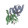 3nonSC  3nooC  3noqC 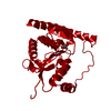 3norC S: Starting model for refinement C: citing same article ( |
|---|---|
| Similar structure data |

-Links
 PDBj
PDBj

- Assembly
| Deposited unit | 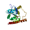
| ||||||||||||
|---|---|---|---|---|---|---|---|---|---|---|---|---|---|
| 1 | 
| ||||||||||||
| Unit cell |
| ||||||||||||
| Components on special symmetry positions |
|
-Components
| #1: Protein | Mass: 24194.672 Da / Num. of mol.: 1 / Mutation: D17E Source method: isolated from a genetically manipulated source Source: (gene. exp.) Pseudomonas fluorescens (bacteria) / Gene: PFL_4109 / Plasmid: pET15b / Production host: |
|---|---|
| #2: Chemical | ChemComp-ACT /   Mass: 59.044 Da / Num. of mol.: 1 / Source method: obtained synthetically / Formula: C2H3O2 Mass: 59.044 Da / Num. of mol.: 1 / Source method: obtained synthetically / Formula: C2H3O2 |
| #3: Water | ChemComp-HOH /  Mass: 18.015 Da / Num. of mol.: 330 / Source method: isolated from a natural source / Formula: H2O Mass: 18.015 Da / Num. of mol.: 330 / Source method: isolated from a natural source / Formula: H2O |
-Experimental details
-Experiment
| Experiment | Method: X-RAY DIFFRACTION / Number of used crystals: 1 |
|---|
- Sample preparation
| Crystal | Density Matthews: 2.48 Å3/Da / Density % sol: 50.47 % |
|---|---|
| Crystal grow | Temperature: 298 K / Method: vapor diffusion, hanging drop / pH: 4.6 Details: 12% PEG 4000, 240 mM ammonium acetate, 100 mM sodium acetate pH=4.6, VAPOR DIFFUSION, HANGING DROP, temperature 298K |
-Data collection
| Diffraction | Mean temperature: 100 K |
|---|---|
| Diffraction source | Source: SYNCHROTRON / Site: APS  / Beamline: 14-BM-C / Wavelength: 0.9 Å / Beamline: 14-BM-C / Wavelength: 0.9 Å |
| Detector | Type: ADSC QUANTUM 315 / Detector: CCD / Date: Oct 24, 2009 / Details: Bent conical Si-mirror (Rh coated) |
| Radiation | Monochromator: Bent Ge(111) monochromator / Protocol: SINGLE WAVELENGTH / Monochromatic (M) / Laue (L): M / Scattering type: x-ray |
| Radiation wavelength | Wavelength: 0.9 Å / Relative weight: 1 |
| Reflection | Resolution: 1.05→37 Å / Num. all: 111571 / Num. obs: 111571 / % possible obs: 99.2 % / Observed criterion σ(F): 0 / Observed criterion σ(I): 0 / Redundancy: 8.6 % / Rmerge(I) obs: 0.08 / Net I/σ(I): 32.9 |
| Reflection shell | Resolution: 1.05→1.09 Å / Redundancy: 5.5 % / Rmerge(I) obs: 0.87 / Mean I/σ(I) obs: 1.9 / Num. unique all: 10915 / % possible all: 98 |
- Processing
| Software |
| |||||||||||||||||||||||||||||||||
|---|---|---|---|---|---|---|---|---|---|---|---|---|---|---|---|---|---|---|---|---|---|---|---|---|---|---|---|---|---|---|---|---|---|---|
| Refinement | Method to determine structure: MOLECULAR REPLACEMENT Starting model: PDB ENTRY 3NON Resolution: 1.05→37 Å / Num. parameters: 19999 / Num. restraintsaints: 26940 / Cross valid method: FREE R / σ(F): 0 / σ(I): 0 / Stereochemistry target values: ENGH AND HUBER Details: ANISOTROPIC SCALING APPLIED BY THE METHOD OF PARKIN, MOEZZI & HOPE, J.APPL.CRYST.28(1995)53-56
| |||||||||||||||||||||||||||||||||
| Solvent computation | Solvent model: BABINET | |||||||||||||||||||||||||||||||||
| Refine analyze | Num. disordered residues: 21 / Occupancy sum hydrogen: 1685.1 / Occupancy sum non hydrogen: 2030.09 | |||||||||||||||||||||||||||||||||
| Refinement step | Cycle: LAST / Resolution: 1.05→37 Å
| |||||||||||||||||||||||||||||||||
| Refine LS restraints |
|
