| Entry | Database: PDB / ID: 6nu8
|
|---|
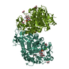
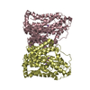
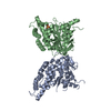
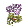
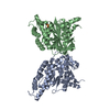
| Title | Structure of sucrose-6-phosphate hydrolase from Lactobacillus gasseri in complex with fructose |
|---|
 Components Components | Sucrose-6-phosphate hydrolase |
|---|
 Keywords Keywords | HYDROLASE / GH32 / glycoside hydrolase / Lactobacillus gasseri / fructose |
|---|
| Function / homology |  Function and homology information Function and homology information
Sucrose-6-phosphate hydrolase / : / Glycoside hydrolase, family 32, active site / Glycosyl hydrolases family 32 active site. / Glycosyl hydrolase family 32, C-terminal / Glycosyl hydrolases family 32 C terminal / Exo-inulinase; domain 1 / Glycoside hydrolase, family 32 / Glycosyl hydrolase family 32, N-terminal / Glycosyl hydrolases family 32 N-terminal domain ...Sucrose-6-phosphate hydrolase / : / Glycoside hydrolase, family 32, active site / Glycosyl hydrolases family 32 active site. / Glycosyl hydrolase family 32, C-terminal / Glycosyl hydrolases family 32 C terminal / Exo-inulinase; domain 1 / Glycoside hydrolase, family 32 / Glycosyl hydrolase family 32, N-terminal / Glycosyl hydrolases family 32 N-terminal domain / Glycosyl hydrolases family 32 / Glycosyl hydrolase domain; family 43 / 5 Propeller / Tachylectin-2; Chain A / Glycosyl hydrolase, five-bladed beta-propellor domain superfamily / Concanavalin A-like lectin/glucanase domain superfamily / Jelly Rolls / Sandwich / Mainly BetaSimilarity search - Domain/homology |
|---|
| Biological species |  Lactobacillus gasseri 224-1 (bacteria) Lactobacillus gasseri 224-1 (bacteria) |
|---|
| Method |  X-RAY DIFFRACTION / SYNCHROTRON / MOLECULAR REPLACEMENT / Resolution: 1.8 Å X-RAY DIFFRACTION / SYNCHROTRON / MOLECULAR REPLACEMENT / Resolution: 1.8 Å |
|---|
 Authors Authors | Lima, M.Z.T. / Muniz, J.R.C. |
|---|
| Funding support |  Brazil, 4items Brazil, 4items | Organization | Grant number | Country |
|---|
| Sao Paulo Research Foundation (FAPESP) | 2017/16291-5 | Brazil | | Brazilian National Council for Scientific and Technological Development (CNPq) | 309767/2015-6 | Brazil | | Brazilian National Council for Scientific and Technological Development (CNPq) | 486546/2013-6 | Brazil | | Brazilian National Council for Scientific and Technological Development (CNPq) | 308865/2018-9 | Brazil |
|
|---|
 Citation Citation | Journal: To Be Published
Title: Structure of sucrose-6-phosphate hydrolase from Lactobacillus gasseri in complex with fructose
Authors: Lima, M.Z.T. / Muniz, J.R.C. |
|---|
| History | | Deposition | Jan 31, 2019 | Deposition site: RCSB / Processing site: RCSB |
|---|
| Revision 1.0 | Feb 5, 2020 | Provider: repository / Type: Initial release |
|---|
| Revision 1.1 | Feb 12, 2020 | Group: Structure summary / Category: struct / Item: _struct.title |
|---|
| Revision 1.2 | Jul 29, 2020 | Group: Data collection / Derived calculations / Structure summary
Category: chem_comp / entity ...chem_comp / entity / pdbx_chem_comp_identifier / pdbx_entity_nonpoly / struct_site / struct_site_gen
Item: _chem_comp.name / _entity.pdbx_description / _pdbx_entity_nonpoly.name
Description: Carbohydrate remediation / Provider: repository / Type: Remediation |
|---|
| Revision 1.3 | Mar 13, 2024 | Group: Data collection / Database references / Structure summary
Category: chem_comp / chem_comp_atom ...chem_comp / chem_comp_atom / chem_comp_bond / database_2
Item: _chem_comp.pdbx_synonyms / _database_2.pdbx_DOI / _database_2.pdbx_database_accession |
|---|
|
|---|
 Movie
Movie Controller

 Yorodumi
 Open data
 Basic information
 Structure visualization
 Downloads & links
 Other downloads


 PDBj
PDBj

 Assembly
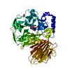

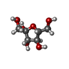
 Type: D-saccharide, beta linking / Mass: 180.156 Da / Num. of mol.: 1
 Mass: 62.068 Da / Num. of mol.: 1 / Source method: obtained synthetically / Formula: C2H6O2
 Mass: 18.015 Da / Num. of mol.: 497 / Source method: isolated from a natural source / Formula: H2O
 Sample preparation
 Processing