 Movie
Movie Controller

[English] 日本語
 Yorodumi
- PDB-4ae2: Crystal structure of Human fibrillar procollagen type III C- prop... -
+ Open data

- Basic information
| Entry | Database: PDB / ID: 4ae2 | ||||||
|---|---|---|---|---|---|---|---|
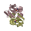
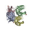
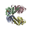
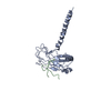
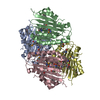
| Title | Crystal structure of Human fibrillar procollagen type III C- propeptide trimer | ||||||
 Components Components | COLLAGEN ALPHA-1(III) CHAIN | ||||||
 Keywords Keywords | STRUCTURAL PROTEIN / FIBRILLAR COLLAGEN / EXTRACELLULAR MATRIX / FIBROSIS | ||||||
| Function / homology |  Function and homology information Function and homology informationcollagen type III trimer / limb joint morphogenesis / aorta smooth muscle tissue morphogenesis / transforming growth factor beta1 production / elastic fiber assembly / Collagen chain trimerization / endochondral bone morphogenesis / peptide cross-linking / platelet-derived growth factor binding / negative regulation of neuron migration ...collagen type III trimer / limb joint morphogenesis / aorta smooth muscle tissue morphogenesis / transforming growth factor beta1 production / elastic fiber assembly / Collagen chain trimerization / endochondral bone morphogenesis / peptide cross-linking / platelet-derived growth factor binding / negative regulation of neuron migration / extracellular matrix structural constituent conferring tensile strength / negative regulation of immune response / layer formation in cerebral cortex / basement membrane organization / Collagen biosynthesis and modifying enzymes / Fibronectin matrix formation / Signaling by PDGF / tissue homeostasis / response to angiotensin / NCAM1 interactions / collagen fibril organization / digestive tract development / Assembly of collagen fibrils and other multimeric structures / MET activates PTK2 signaling / Scavenging by Class A Receptors / extracellular matrix structural constituent / skin development / Syndecan interactions / positive regulation of Rho protein signal transduction / SMAD binding / Collagen degradation / Non-integrin membrane-ECM interactions / chondrocyte differentiation / ECM proteoglycans / Integrin cell surface interactions / supramolecular fiber organization / response to cytokine / transforming growth factor beta receptor signaling pathway / lung development / cell-matrix adhesion / Developmental Lineage of Pancreatic Ductal Cells / integrin-mediated signaling pathway / wound healing / cellular response to amino acid stimulus / platelet activation / response to radiation / cerebral cortex development / integrin binding / multicellular organism growth / neuron migration / Immunoregulatory interactions between a Lymphoid and a non-Lymphoid cell / heart development / extracellular matrix / protease binding / fibroblast proliferation / in utero embryonic development / endoplasmic reticulum lumen / : / extracellular region / metal ion binding Similarity search - Function | ||||||
| Biological species |  HOMO SAPIENS (human) HOMO SAPIENS (human) | ||||||
| Method |  X-RAY DIFFRACTION / SYNCHROTRON / MOLECULAR REPLACEMENT / Resolution: 1.68 Å X-RAY DIFFRACTION / SYNCHROTRON / MOLECULAR REPLACEMENT / Resolution: 1.68 Å | ||||||
 Authors Authors | Bourhis, J.M. / Mariano, N. / Zhao, Y. / Harlos, K. / Jones, E.Y. / Moali, C. / Aghajari, N. / Hulmes, D.J. | ||||||
 Citation Citation | Journal: Nat.Struct.Mol.Biol. / Year: 2012 Title: Structural Basis of Fibrillar Collagen Trimerization and Related Genetic Disorders. Authors: Bourhis, J.M. / Mariano, N. / Zhao, Y. / Harlos, K. / Exposito, J.Y. / Jones, E.Y. / Moali, C. / Aghajari, N. / Hulmes, D.J. #1: Journal: Acta Crystallogr.,Sect.F / Year: 2012 Title: Production and Crystallization of the C-Propeptide Trimer from Human Procollagen III. Authors: Bourhis, J.M. / Mariano, N. / Zhao, Y. / Walter, T.S. / El Omari, K. / Delolme, F. / Moali, C. / Hulmes, D.J. / Aghajari, N. | ||||||
| History |
|
- Structure visualization
| Structure viewer | Molecule: MolmilJmol/JSmol |
|---|
- Downloads & links
-Download
| PDBx/mmCIF format | 4ae2.cif.gz | 280.2 KB | Display | PDBx/mmCIF format |
|---|---|---|---|---|
| PDB format | pdb4ae2.ent.gz | 225.6 KB | Display | PDB format |
| PDBx/mmJSON format | 4ae2.json.gz | Tree view | PDBx/mmJSON format | |
| Others |  Other downloads Other downloads |
-Validation report
| Arichive directory | https://data.pdbj.org/pub/pdb/validation_reports/ae/4ae2ftp://data.pdbj.org/pub/pdb/validation_reports/ae/4ae2 | HTTPS FTP |
|---|
-Related structure data


-Links
 PDBj
PDBj


- Assembly
| Deposited unit | 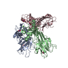
| ||||||||
|---|---|---|---|---|---|---|---|---|---|
| 1 |
| ||||||||
| Unit cell |
|
-Components
| #1: Protein | Mass: 28815.340 Da / Num. of mol.: 3 Fragment: CPROPEPTIDE OF PROCOLLAGEN III, RESIDUES 1222-1466 Mutation: YES Source method: isolated from a genetically manipulated source Source: (gene. exp.) HOMO SAPIENS (human) / Plasmid: PHLSEC / Cell line (production host): HEK293T / Production host: HOMO SAPIENS (human) / References: UniProt: P02461#2: Chemical |   Mass: 40.078 Da / Num. of mol.: 3 / Source method: obtained synthetically / Formula: Ca Mass: 40.078 Da / Num. of mol.: 3 / Source method: obtained synthetically / Formula: Ca#3: Chemical | ChemComp-NO3 /   Mass: 62.005 Da / Num. of mol.: 5 / Source method: obtained synthetically / Formula: NO3 Mass: 62.005 Da / Num. of mol.: 5 / Source method: obtained synthetically / Formula: NO3#4: Water | ChemComp-HOH / |  Mass: 18.015 Da / Num. of mol.: 398 / Source method: isolated from a natural source / Formula: H2O Mass: 18.015 Da / Num. of mol.: 398 / Source method: isolated from a natural source / Formula: H2OHas protein modification | Y | |
|---|
-Experimental details
-Experiment
| Experiment | Method: X-RAY DIFFRACTION / Number of used crystals: 1 |
|---|
- Sample preparation
| Crystal | Density Matthews: 2 Å3/Da / Density % sol: 39.9 % / Description: NONE |
|---|---|
| Crystal grow | pH: 6.5 Details: 20% PEG 3350, 0.1 M BIS TRIS PROPANE PH 6.5, POTASSIUM NITRATE 0.2 M |
-Data collection
| Diffraction | Mean temperature: 100 K |
|---|---|
| Diffraction source | Source: SYNCHROTRON / Site: Diamond  / Beamline: I03 / Wavelength: 0.976 / Beamline: I03 / Wavelength: 0.976 |
| Detector | Type: ADSC CCD / Detector: CCD / Date: May 16, 2011 |
| Radiation | Protocol: SINGLE WAVELENGTH / Monochromatic (M) / Laue (L): M / Scattering type: x-ray |
| Radiation wavelength | Wavelength: 0.976 Å / Relative weight: 1 |
| Reflection | Resolution: 1.7→61.2 Å / Num. obs: 78019 / % possible obs: 96.6 % / Observed criterion σ(I): 2 / Redundancy: 3.5 % / Rmerge(I) obs: 0.07 / Net I/σ(I): 9.6 |
| Reflection shell | Resolution: 1.7→61 Å / Redundancy: 3.6 % / Rmerge(I) obs: 0.4 / Mean I/σ(I) obs: 3 / % possible all: 96.2 |
- Processing
| Software |
| ||||||||||||||||||||||||||||||||||||||||||||||||||||||||||||||||||||||||||||||||||||||||||||||||||||||||||||||||||||||||||||||||||||||||||||||||||||||||||||||||||||||||||||||||||||||
|---|---|---|---|---|---|---|---|---|---|---|---|---|---|---|---|---|---|---|---|---|---|---|---|---|---|---|---|---|---|---|---|---|---|---|---|---|---|---|---|---|---|---|---|---|---|---|---|---|---|---|---|---|---|---|---|---|---|---|---|---|---|---|---|---|---|---|---|---|---|---|---|---|---|---|---|---|---|---|---|---|---|---|---|---|---|---|---|---|---|---|---|---|---|---|---|---|---|---|---|---|---|---|---|---|---|---|---|---|---|---|---|---|---|---|---|---|---|---|---|---|---|---|---|---|---|---|---|---|---|---|---|---|---|---|---|---|---|---|---|---|---|---|---|---|---|---|---|---|---|---|---|---|---|---|---|---|---|---|---|---|---|---|---|---|---|---|---|---|---|---|---|---|---|---|---|---|---|---|---|---|---|---|---|
| Refinement | Method to determine structure: MOLECULAR REPLACEMENT / Resolution: 1.68→61.27 Å / Cor.coef. Fo:Fc: 0.964 / Cor.coef. Fo:Fc free: 0.946 / SU B: 4.44 / SU ML: 0.067 / Cross valid method: THROUGHOUT / ESU R: 0.13 / ESU R Free: 0.1 / Stereochemistry target values: MAXIMUM LIKELIHOOD / Details: HYDROGENS HAVE BEEN ADDED IN THE RIDING POSITIONS.
| ||||||||||||||||||||||||||||||||||||||||||||||||||||||||||||||||||||||||||||||||||||||||||||||||||||||||||||||||||||||||||||||||||||||||||||||||||||||||||||||||||||||||||||||||||||||
| Solvent computation | Ion probe radii: 0.8 Å / Shrinkage radii: 0.8 Å / VDW probe radii: 1.2 Å / Solvent model: MASK | ||||||||||||||||||||||||||||||||||||||||||||||||||||||||||||||||||||||||||||||||||||||||||||||||||||||||||||||||||||||||||||||||||||||||||||||||||||||||||||||||||||||||||||||||||||||
| Displacement parameters | Biso mean: 21.855 Å2
| ||||||||||||||||||||||||||||||||||||||||||||||||||||||||||||||||||||||||||||||||||||||||||||||||||||||||||||||||||||||||||||||||||||||||||||||||||||||||||||||||||||||||||||||||||||||
| Refinement step | Cycle: LAST / Resolution: 1.68→61.27 Å
| ||||||||||||||||||||||||||||||||||||||||||||||||||||||||||||||||||||||||||||||||||||||||||||||||||||||||||||||||||||||||||||||||||||||||||||||||||||||||||||||||||||||||||||||||||||||
| Refine LS restraints |
|