 Movie
Movie Controller


+ Open data

- Basic information
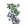
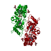
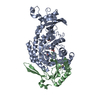
| Entry | Database: PDB / ID: 4aam | ||||||
|---|---|---|---|---|---|---|---|
| Title | MacA wild-type mixed-valence | ||||||
 Components Components | CYTOCHROME C551 PEROXIDASE | ||||||
 Keywords Keywords | OXIDOREDUCTASE / MULTIHEME CYTOCHROMES / CONFORMATIONAL REARRANGEMENT | ||||||
| Function / homology |  Function and homology information Function and homology informationcytochrome-c peroxidase / cytochrome-c peroxidase activity / periplasmic space / electron transfer activity / heme binding / metal ion binding Similarity search - Function | ||||||
| Biological species |  GEOBACTER SULFURREDUCENS (bacteria) GEOBACTER SULFURREDUCENS (bacteria) | ||||||
| Method |  X-RAY DIFFRACTION / SYNCHROTRON / MOLECULAR REPLACEMENT / Resolution: 2.17 Å X-RAY DIFFRACTION / SYNCHROTRON / MOLECULAR REPLACEMENT / Resolution: 2.17 Å | ||||||
 Authors Authors | Seidel, J. | ||||||
 Citation Citation | Journal: Biochemistry / Year: 2012 Title: Maca is a Second Cytochrome C Peroxidase of Geobacter Sulfurreducens. Authors: Seidel, J. / Hoffmann, M. / Ellis, K.E. / Seidel, A. / Spatzal, T. / Gerhardt, S. / Elliott, S.J. / Einsle, O. | ||||||
| History |
|
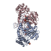
- Structure visualization
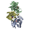
| Structure viewer | Molecule: MolmilJmol/JSmol |
|---|
- Downloads & links
-Download
| PDBx/mmCIF format | 4aam.cif.gz | 145 KB | Display | PDBx/mmCIF format |
|---|---|---|---|---|
| PDB format | pdb4aam.ent.gz | 111.8 KB | Display | PDB format |
| PDBx/mmJSON format | 4aam.json.gz | Tree view | PDBx/mmJSON format | |
| Others |  Other downloads Other downloads |
-Validation report
| Summary document | 4aam_validation.pdf.gz | 1.1 MB | Display | wwPDB validaton report |
|---|---|---|---|---|
| Full document | 4aam_full_validation.pdf.gz | 1.1 MB | Display | |
| Data in XML | 4aam_validation.xml.gz | 18.2 KB | Display | |
| Data in CIF | 4aam_validation.cif.gz | 27.3 KB | Display | |
| Arichive directory | https://data.pdbj.org/pub/pdb/validation_reports/aa/4aamftp://data.pdbj.org/pub/pdb/validation_reports/aa/4aam | HTTPS FTP |
-Related structure data
| Related structure data | 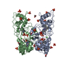 4aalC  4aanC  4aaoC 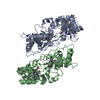 3hq6S C: citing same article ( S: Starting model for refinement |
|---|---|
| Similar structure data |


-Links
 PDBj
PDBj


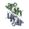
- Assembly
| Deposited unit | 
| ||||||||
|---|---|---|---|---|---|---|---|---|---|
| 1 | 
| ||||||||
| Unit cell |
|
-Components
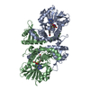
| #1: Protein | Mass: 36814.848 Da / Num. of mol.: 1 / Fragment: RESIDUES 23-346 Source method: isolated from a genetically manipulated source Source: (gene. exp.) GEOBACTER SULFURREDUCENS (bacteria) / Strain: PCA / Description: DSM / Plasmid: PETSN22 / Production host: | ||||||||
|---|---|---|---|---|---|---|---|---|---|
| #2: Chemical |   Mass: 618.503 Da / Num. of mol.: 2 / Source method: obtained synthetically / Formula: C34H34FeN4O4 Mass: 618.503 Da / Num. of mol.: 2 / Source method: obtained synthetically / Formula: C34H34FeN4O4#3: Chemical | ChemComp-CA / |   Mass: 40.078 Da / Num. of mol.: 1 / Source method: obtained synthetically / Formula: Ca Mass: 40.078 Da / Num. of mol.: 1 / Source method: obtained synthetically / Formula: Ca#4: Chemical |   Mass: 96.063 Da / Num. of mol.: 2 / Source method: obtained synthetically / Formula: SO4 Mass: 96.063 Da / Num. of mol.: 2 / Source method: obtained synthetically / Formula: SO4#5: Water | ChemComp-HOH / |  Mass: 18.015 Da / Num. of mol.: 320 / Source method: isolated from a natural source / Formula: H2O Mass: 18.015 Da / Num. of mol.: 320 / Source method: isolated from a natural source / Formula: H2OHas protein modification | Y | |
-Experimental details
-Experiment
| Experiment | Method: X-RAY DIFFRACTION / Number of used crystals: 1 |
|---|
- Sample preparation
| Crystal | Density Matthews: 2.51 Å3/Da / Density % sol: 51.05 % / Description: NONE |
|---|---|
| Crystal grow | pH: 7.5 Details: 0.1 M NAHEPES PH 7.5, 0.2 M AMMONIUM ACETATE, 25% POLYETHYLENE GLYCOL 3350 |
-Data collection
| Diffraction | Mean temperature: 100 K |
|---|---|
| Diffraction source | Source: SYNCHROTRON / Site: SLS  / Beamline: X06DA / Wavelength: 1 / Beamline: X06DA / Wavelength: 1 |
| Detector | Type: MARMOSAIC 225 mm CCD / Detector: CCD / Date: Dec 10, 2009 |
| Radiation | Protocol: SINGLE WAVELENGTH / Monochromatic (M) / Laue (L): M / Scattering type: x-ray |
| Radiation wavelength | Wavelength: 1 Å / Relative weight: 1 |
| Reflection | Resolution: 2.2→40.1 Å / Num. obs: 18659 / % possible obs: 99.9 % / Observed criterion σ(I): 0 / Redundancy: 3.5 % / Rmerge(I) obs: 0.12 / Net I/σ(I): 6.4 |
| Reflection shell | Resolution: 2.2→2.3 Å / Redundancy: 3.5 % / Rmerge(I) obs: 0.23 / Mean I/σ(I) obs: 4 / % possible all: 99.9 |
- Processing
| Software |
| ||||||||||||||||||||||||||||||||||||||||||||||||||||||||||||||||||||||||||||||||||||||||||||||||||||||||||||||||||||||||||||||||||||||||||||||||||||||||||||||||||||||||||||||||||||||
|---|---|---|---|---|---|---|---|---|---|---|---|---|---|---|---|---|---|---|---|---|---|---|---|---|---|---|---|---|---|---|---|---|---|---|---|---|---|---|---|---|---|---|---|---|---|---|---|---|---|---|---|---|---|---|---|---|---|---|---|---|---|---|---|---|---|---|---|---|---|---|---|---|---|---|---|---|---|---|---|---|---|---|---|---|---|---|---|---|---|---|---|---|---|---|---|---|---|---|---|---|---|---|---|---|---|---|---|---|---|---|---|---|---|---|---|---|---|---|---|---|---|---|---|---|---|---|---|---|---|---|---|---|---|---|---|---|---|---|---|---|---|---|---|---|---|---|---|---|---|---|---|---|---|---|---|---|---|---|---|---|---|---|---|---|---|---|---|---|---|---|---|---|---|---|---|---|---|---|---|---|---|---|---|
| Refinement | Method to determine structure: MOLECULAR REPLACEMENT Starting model: PDB ENTRY 3HQ6 Resolution: 2.17→77.82 Å / Cor.coef. Fo:Fc: 0.945 / Cor.coef. Fo:Fc free: 0.883 / SU B: 9.813 / SU ML: 0.14 / Cross valid method: THROUGHOUT / ESU R: 0.244 / ESU R Free: 0.213 / Stereochemistry target values: MAXIMUM LIKELIHOOD Details: HYDROGENS HAVE BEEN ADDED IN THE RIDING POSITIONS. HYDROGENS HAVE BEEN USED IF PRESENT IN THE INPUT.
| ||||||||||||||||||||||||||||||||||||||||||||||||||||||||||||||||||||||||||||||||||||||||||||||||||||||||||||||||||||||||||||||||||||||||||||||||||||||||||||||||||||||||||||||||||||||
| Solvent computation | Ion probe radii: 0.8 Å / Shrinkage radii: 0.8 Å / VDW probe radii: 1.2 Å / Solvent model: MASK | ||||||||||||||||||||||||||||||||||||||||||||||||||||||||||||||||||||||||||||||||||||||||||||||||||||||||||||||||||||||||||||||||||||||||||||||||||||||||||||||||||||||||||||||||||||||
| Displacement parameters | Biso mean: 17.593 Å2
| ||||||||||||||||||||||||||||||||||||||||||||||||||||||||||||||||||||||||||||||||||||||||||||||||||||||||||||||||||||||||||||||||||||||||||||||||||||||||||||||||||||||||||||||||||||||
| Refinement step | Cycle: LAST / Resolution: 2.17→77.82 Å
| ||||||||||||||||||||||||||||||||||||||||||||||||||||||||||||||||||||||||||||||||||||||||||||||||||||||||||||||||||||||||||||||||||||||||||||||||||||||||||||||||||||||||||||||||||||||
| Refine LS restraints |
|