 Movie
Movie Controller

[English] 日本語
 Yorodumi
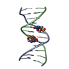
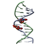
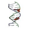
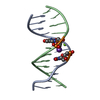
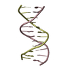
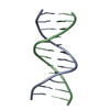
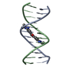
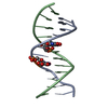
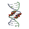
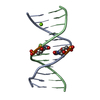
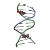
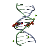
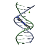
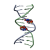
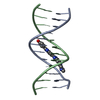
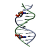
- PDB-455d: A6/A18 INTER-STRAND DITHIOBIS(PROPANE)-CROSSLINKED DODECAMER (CGC... -
+ Open data

- Basic information
| Entry | Database: PDB / ID: 455d | ||||||||||||||||||
|---|---|---|---|---|---|---|---|---|---|---|---|---|---|---|---|---|---|---|---|
| Title | A6/A18 INTER-STRAND DITHIOBIS(PROPANE)-CROSSLINKED DODECAMER (CGCGAATTCGCG)2 | ||||||||||||||||||
 Components Components | DNA (5'-D(* Keywords KeywordsDNA / INTER-STRAND CROSSLINKING / DODECAMER CGCGAATTCGCG/CGCGAATTCGCG / DEOXYRIBONUCLEIC ACID | Function / homology | DITHIOBIS-(PROPANE) / DNA / DNA (> 10) |  Function and homology information Function and homology informationMethod |  X-RAY DIFFRACTION / MOLECULAR REPLACEMEN / Resolution: 1.43 Å X-RAY DIFFRACTION / MOLECULAR REPLACEMEN / Resolution: 1.43 Å  Authors AuthorsChiu, T.K. / Kaczor-Grzeskowiak, M. / Dickerson, R.E. |  CitationJournal: J.Mol.Biol. / Year: 1999 CitationJournal: J.Mol.Biol. / Year: 1999Title: Absence of minor groove monovalent cations in the crosslinked dodecamer C-G-C-G-A-A-T-T-C-G-C-G. Authors: Chiu, T.K. / Kaczor-Grzeskowiak, M. / Dickerson, R.E. History |
|
- Structure visualization
| Structure viewer | Molecule: MolmilJmol/JSmol |
|---|
- Downloads & links
-Download
| PDBx/mmCIF format | 455d.cif.gz | 27.1 KB | Display | PDBx/mmCIF format |
|---|---|---|---|---|
| PDB format | pdb455d.ent.gz | 17.4 KB | Display | PDB format |
| PDBx/mmJSON format | 455d.json.gz | Tree view | PDBx/mmJSON format | |
| Others |  Other downloads Other downloads |
-Validation report
| Arichive directory | https://data.pdbj.org/pub/pdb/validation_reports/55/455dftp://data.pdbj.org/pub/pdb/validation_reports/55/455d | HTTPS FTP |
|---|
-Related structure data
| Similar structure data |
|---|


-Links
 PDBj
PDBj


- Assembly
| Deposited unit | 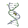
| ||||||||||
|---|---|---|---|---|---|---|---|---|---|---|---|
| 1 |
| ||||||||||
| Unit cell |
|
-Components
| #1: DNA chain | Mass: 3663.392 Da / Num. of mol.: 2 / Source method: obtained synthetically / Details: COMPLEXED WITH THIOPROPANE #2: Chemical |   Mass: 24.305 Da / Num. of mol.: 3 / Source method: obtained synthetically / Formula: Mg Mass: 24.305 Da / Num. of mol.: 3 / Source method: obtained synthetically / Formula: Mg#3: Chemical | ChemComp-SSP / | 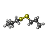  Mass: 150.305 Da / Num. of mol.: 1 / Source method: obtained synthetically / Formula: C6H14S2 Mass: 150.305 Da / Num. of mol.: 1 / Source method: obtained synthetically / Formula: C6H14S2#4: Water | ChemComp-HOH / |  Mass: 18.015 Da / Num. of mol.: 150 / Source method: isolated from a natural source / Formula: H2O Mass: 18.015 Da / Num. of mol.: 150 / Source method: isolated from a natural source / Formula: H2O |
|---|
-Experimental details
-Experiment
| Experiment | Method: X-RAY DIFFRACTION / Number of used crystals: 1 |
|---|
- Sample preparation
| Crystal | Density Matthews: 1.78 Å3/Da / Density % sol: 30.5 % | ||||||||||||||||||||||||||||||||||||||||||
|---|---|---|---|---|---|---|---|---|---|---|---|---|---|---|---|---|---|---|---|---|---|---|---|---|---|---|---|---|---|---|---|---|---|---|---|---|---|---|---|---|---|---|---|
| Crystal grow | Temperature: 277 K / Method: vapor diffusion, hanging drop / Details: VAPOR DIFFUSION, HANGING DROP, temperature 277K | ||||||||||||||||||||||||||||||||||||||||||
| Components of the solutions |
| ||||||||||||||||||||||||||||||||||||||||||
| Crystal | *PLUS | ||||||||||||||||||||||||||||||||||||||||||
| Crystal grow | *PLUS Temperature: 4 ℃ / pH: 7 / Method: vapor diffusion, sitting drop | ||||||||||||||||||||||||||||||||||||||||||
| Components of the solutions | *PLUS
|
-Data collection
| Diffraction | Mean temperature: 100 K |
|---|---|
| Diffraction source | Source: ROTATING ANODE / Type: RIGAKU / Wavelength: 1.5418 |
| Detector | Type: RIGAKU RAXIS IV / Detector: IMAGE PLATE |
| Radiation | Monochromator: MIRRORS / Protocol: SINGLE WAVELENGTH / Monochromatic (M) / Laue (L): M / Scattering type: x-ray |
| Radiation wavelength | Wavelength: 1.5418 Å / Relative weight: 1 |
| Reflection | Resolution: 1.43→8 Å / Num. obs: 11200 / % possible obs: 89.4 % / Observed criterion σ(I): -3 / Redundancy: 9.49 % / Biso Wilson estimate: 17.85 Å2 / Rmerge(I) obs: 0.037 / Net I/σ(I): 38.1 |
| Reflection shell | Resolution: 1.43→1.54 Å / Redundancy: 6.73 % / Rmerge(I) obs: 0.156 / Mean I/σ(I) obs: 10.9 / % possible all: 80.8 |
| Reflection | *PLUS Lowest resolution: 8 Å / Observed criterion σ(I): 2 / Num. measured all: 106309 |
| Reflection shell | *PLUS % possible obs: 80.8 % / Mean I/σ(I) obs: 10.93 |
- Processing
| Software |
| ||||||||||||||||||||||||||||||||||||||||||||||||||||||||||||
|---|---|---|---|---|---|---|---|---|---|---|---|---|---|---|---|---|---|---|---|---|---|---|---|---|---|---|---|---|---|---|---|---|---|---|---|---|---|---|---|---|---|---|---|---|---|---|---|---|---|---|---|---|---|---|---|---|---|---|---|---|---|
| Refinement | Method to determine structure: MOLECULAR REPLACEMEN Starting model: IDEAL B-DNA Resolution: 1.43→8 Å / Cross valid method: THROUGHOUT / σ(F): 2 Details: STARTING MODEL IS IDEAL B-DNA RMS-FITTED ONTO COORDINATES OF ORIGINAL DREW STRUCTURE (1BNA)
| ||||||||||||||||||||||||||||||||||||||||||||||||||||||||||||
| Displacement parameters | Biso mean: 13.56 Å2
| ||||||||||||||||||||||||||||||||||||||||||||||||||||||||||||
| Refinement step | Cycle: LAST / Resolution: 1.43→8 Å
| ||||||||||||||||||||||||||||||||||||||||||||||||||||||||||||
| Refine LS restraints |
| ||||||||||||||||||||||||||||||||||||||||||||||||||||||||||||
| Software | *PLUS Name: X-PLOR / Version: 3.843 / Classification: refinement | ||||||||||||||||||||||||||||||||||||||||||||||||||||||||||||
| Refinement | *PLUS Lowest resolution: 8 Å / σ(F): 2 / % reflection Rfree: 10 % | ||||||||||||||||||||||||||||||||||||||||||||||||||||||||||||
| Solvent computation | *PLUS | ||||||||||||||||||||||||||||||||||||||||||||||||||||||||||||
| Displacement parameters | *PLUS | ||||||||||||||||||||||||||||||||||||||||||||||||||||||||||||
| Refine LS restraints | *PLUS
|
