 Movie
Movie Controller

[English] 日本語
 Yorodumi
- PDB-3zse: 3D Structure of a thermophilic family GH11 xylanase from Thermobi... -
+ Open data

- Basic information
| Entry | Database: PDB / ID: 3zse | |||||||||
|---|---|---|---|---|---|---|---|---|---|---|
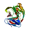
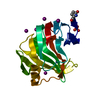
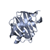
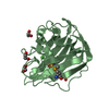
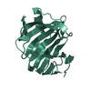
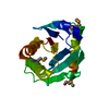
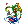
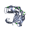
| Title | 3D Structure of a thermophilic family GH11 xylanase from Thermobifida fusca | |||||||||
 Components Components | ENDO-1,4-BETA-XYLANASE | |||||||||
 Keywords Keywords | HYDROLASE / GLYCOSIDE HYDROLASE / PLANT CELL WALL | |||||||||
| Function / homology |  Function and homology information Function and homology informationendo-1,4-beta-xylanase / endo-1,4-beta-xylanase activity / xylan catabolic process / polysaccharide binding Similarity search - Function | |||||||||
| Biological species |   THERMOBIFIDA FUSCA (bacteria) THERMOBIFIDA FUSCA (bacteria) | |||||||||
| Method |  X-RAY DIFFRACTION / SYNCHROTRON / MOLECULAR REPLACEMENT / Resolution: 1.78 Å X-RAY DIFFRACTION / SYNCHROTRON / MOLECULAR REPLACEMENT / Resolution: 1.78 Å | |||||||||
 Authors Authors | Lammerts van Bueren, A. / Otani, S. / Friis, E.P. / S Wilson, K. / Davies, G.J. | |||||||||
 Citation Citation | Journal: Acta Crystallogr.,Sect.F / Year: 2012 Title: Three-Dimensional Structure of a Thermophilic Family Gh11 Xylanase from Thermobifida Fusca. Authors: Lammerts Van Bueren, A. / Otani, S. / Friis, E.P. / Wilson, K.S. / Davies, G.J. | |||||||||
| History |
|
- Structure visualization
| Structure viewer | Molecule: MolmilJmol/JSmol |
|---|
- Downloads & links
-Download
| PDBx/mmCIF format | 3zse.cif.gz | 89.1 KB | Display | PDBx/mmCIF format |
|---|---|---|---|---|
| PDB format | pdb3zse.ent.gz | 66.3 KB | Display | PDB format |
| PDBx/mmJSON format | 3zse.json.gz | Tree view | PDBx/mmJSON format | |
| Others |  Other downloads Other downloads |
-Validation report
| Arichive directory | https://data.pdbj.org/pub/pdb/validation_reports/zs/3zseftp://data.pdbj.org/pub/pdb/validation_reports/zs/3zse | HTTPS FTP |
|---|
-Related structure data
| Related structure data |  1qh6S S: Starting model for refinement |
|---|---|
| Similar structure data |


-Links
 PDBj
PDBj

- Assembly
| Deposited unit | 
| ||||||||
|---|---|---|---|---|---|---|---|---|---|
| 1 |
| ||||||||
| Unit cell |
|
-Components
| #1: Protein | Mass: 22251.018 Da / Num. of mol.: 1 / Fragment: CATALYTIC DOMAIN, RESIDUES 43-236 Source method: isolated from a genetically manipulated source Details: COVALENT GLYCOSYL-ENZYME INTERMEDIATE TO GLU127 / Source: (gene. exp.) THERMOBIFIDA FUSCA (bacteria) / Production host: | ||||
|---|---|---|---|---|---|
| #2: Polysaccharide | beta-D-xylopyranose-(1-4)-2-deoxy-2-fluoro-alpha-D-xylopyranose Source method: isolated from a genetically manipulated source | ||||
| #3: Chemical | ChemComp-EDO /   Mass: 62.068 Da / Num. of mol.: 5 / Source method: obtained synthetically / Formula: C2H6O2 Mass: 62.068 Da / Num. of mol.: 5 / Source method: obtained synthetically / Formula: C2H6O2#4: Water | ChemComp-HOH / |  Mass: 18.015 Da / Num. of mol.: 146 / Source method: isolated from a natural source / Formula: H2O Mass: 18.015 Da / Num. of mol.: 146 / Source method: isolated from a natural source / Formula: H2OHas protein modification | Y | |
-Experimental details
-Experiment
| Experiment | Method: X-RAY DIFFRACTION / Number of used crystals: 1 |
|---|
- Sample preparation
| Crystal | Density Matthews: 1.9 Å3/Da / Density % sol: 35 % / Description: NONE |
|---|---|
| Crystal grow | pH: 4.6 Details: 200MM POTASSIUM CITRATE, 0.2M CACL2, 15% PEG3350, 8% PEG550MME, 0.1M SODIUM ACETATE PH4.6. PROTEIN AT 8 MG/ML |
-Data collection
| Diffraction | Mean temperature: 100 K |
|---|---|
| Diffraction source | Source: SYNCHROTRON / Site: Diamond  / Beamline: I04 / Wavelength: 0.976 / Beamline: I04 / Wavelength: 0.976 |
| Detector | Type: ADSC CCD / Detector: CCD |
| Radiation | Protocol: SINGLE WAVELENGTH / Monochromatic (M) / Laue (L): M / Scattering type: x-ray |
| Radiation wavelength | Wavelength: 0.976 Å / Relative weight: 1 |
| Reflection | Resolution: 1.78→45 Å / Num. obs: 15691 / % possible obs: 99.9 % / Observed criterion σ(I): 0 / Redundancy: 5 % / Rmerge(I) obs: 0.15 / Net I/σ(I): 6.4 |
| Reflection shell | Resolution: 1.78→1.88 Å / Redundancy: 5 % / Rmerge(I) obs: 0.5 / Mean I/σ(I) obs: 2.2 / % possible all: 99.9 |
- Processing
| Software |
| ||||||||||||||||||||||||||||||||||||||||||||||||||||||||||||||||||||||||||||||||||||||||||||||||||||||||||||||||||||||||||||||||||||||||||||||||||||||||||||||||||||||||||||||||||||||
|---|---|---|---|---|---|---|---|---|---|---|---|---|---|---|---|---|---|---|---|---|---|---|---|---|---|---|---|---|---|---|---|---|---|---|---|---|---|---|---|---|---|---|---|---|---|---|---|---|---|---|---|---|---|---|---|---|---|---|---|---|---|---|---|---|---|---|---|---|---|---|---|---|---|---|---|---|---|---|---|---|---|---|---|---|---|---|---|---|---|---|---|---|---|---|---|---|---|---|---|---|---|---|---|---|---|---|---|---|---|---|---|---|---|---|---|---|---|---|---|---|---|---|---|---|---|---|---|---|---|---|---|---|---|---|---|---|---|---|---|---|---|---|---|---|---|---|---|---|---|---|---|---|---|---|---|---|---|---|---|---|---|---|---|---|---|---|---|---|---|---|---|---|---|---|---|---|---|---|---|---|---|---|---|
| Refinement | Method to determine structure: MOLECULAR REPLACEMENT Starting model: PDB ENTRY 1QH6 Resolution: 1.78→37.29 Å / Cor.coef. Fo:Fc: 0.948 / Cor.coef. Fo:Fc free: 0.929 / SU B: 6.254 / SU ML: 0.101 / Cross valid method: THROUGHOUT / ESU R: 0.158 / ESU R Free: 0.147 / Stereochemistry target values: MAXIMUM LIKELIHOOD Details: HYDROGENS HAVE BEEN ADDED IN THE RIDING POSITIONS. HYDROGENS HAVE BEEN USED IF PRESENT IN THE INPUT U VALUES WITH TLS ADDED
| ||||||||||||||||||||||||||||||||||||||||||||||||||||||||||||||||||||||||||||||||||||||||||||||||||||||||||||||||||||||||||||||||||||||||||||||||||||||||||||||||||||||||||||||||||||||
| Solvent computation | Ion probe radii: 0.8 Å / Shrinkage radii: 0.8 Å / VDW probe radii: 1.2 Å / Solvent model: MASK | ||||||||||||||||||||||||||||||||||||||||||||||||||||||||||||||||||||||||||||||||||||||||||||||||||||||||||||||||||||||||||||||||||||||||||||||||||||||||||||||||||||||||||||||||||||||
| Displacement parameters | Biso mean: 15.947 Å2
| ||||||||||||||||||||||||||||||||||||||||||||||||||||||||||||||||||||||||||||||||||||||||||||||||||||||||||||||||||||||||||||||||||||||||||||||||||||||||||||||||||||||||||||||||||||||
| Refinement step | Cycle: LAST / Resolution: 1.78→37.29 Å
| ||||||||||||||||||||||||||||||||||||||||||||||||||||||||||||||||||||||||||||||||||||||||||||||||||||||||||||||||||||||||||||||||||||||||||||||||||||||||||||||||||||||||||||||||||||||
| Refine LS restraints |
|