 Movie
Movie Controller

[English] 日本語
 Yorodumi
- PDB-3zla: Crystal structure of the nucleocapsid protein from Bunyamwera vir... -
+ Open data

- Basic information
| Entry | Database: PDB / ID: 3zla | ||||||
|---|---|---|---|---|---|---|---|
| Title | Crystal structure of the nucleocapsid protein from Bunyamwera virus bound to RNA | ||||||
 Components Components |
| ||||||
 Keywords Keywords | VIRAL PROTEIN/RNA / VIRAL PROTEIN-RNA COMPLEX | ||||||
| Function / homology |  Function and homology information Function and homology informationhelical viral capsid / viral nucleocapsid / ribonucleoprotein complex / symbiont-mediated suppression of host gene expression / RNA binding Similarity search - Function | ||||||
| Biological species |  BUNYAMWERA VIRUS BUNYAMWERA VIRUS | ||||||
| Method |  X-RAY DIFFRACTION / SYNCHROTRON / MOLECULAR REPLACEMENT / Resolution: 3.2 Å X-RAY DIFFRACTION / SYNCHROTRON / MOLECULAR REPLACEMENT / Resolution: 3.2 Å | ||||||
 Authors Authors | Ariza, A. / Tanner, S.J. / Walter, C.T. / Dent, K.C. / Shepherd, D.A. / Wu, W. / Matthews, S.V. / Hiscox, J.A. / Green, T.J. / Luo, M. ...Ariza, A. / Tanner, S.J. / Walter, C.T. / Dent, K.C. / Shepherd, D.A. / Wu, W. / Matthews, S.V. / Hiscox, J.A. / Green, T.J. / Luo, M. / Elliot, R.M. / Ashcroft, A.E. / Stonehouse, N.J. / Ranson, N.A. / Barr, J.N. / Edwards, T.A. | ||||||
 Citation Citation | Journal: Nucleic Acids Res. / Year: 2013 Title: Nucleocapsid Protein Structures from Orthobunyaviruses Reveal Insight Into Ribonucleoprotein Architecture and RNA Polymerization. Authors: Ariza, A. / Tanner, S.J. / Walter, C.T. / Dent, K.C. / Shepherd, D.A. / Wu, W. / Matthews, S.V. / Hiscox, J.A. / Green, T.J. / Luo, M. / Elliott, R.M. / Fooks, A.R. / Ashcroft, A.E. / ...Authors: Ariza, A. / Tanner, S.J. / Walter, C.T. / Dent, K.C. / Shepherd, D.A. / Wu, W. / Matthews, S.V. / Hiscox, J.A. / Green, T.J. / Luo, M. / Elliott, R.M. / Fooks, A.R. / Ashcroft, A.E. / Stonehouse, N.J. / Ranson, N.A. / Barr, J.N. / Edwards, T.A. | ||||||
| History |
|
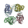
- Structure visualization
| Structure viewer | Molecule: MolmilJmol/JSmol |
|---|
- Downloads & links
-Download
| PDBx/mmCIF format | 3zla.cif.gz | 404.9 KB | Display | PDBx/mmCIF format |
|---|---|---|---|---|
| PDB format | pdb3zla.ent.gz | 336.8 KB | Display | PDB format |
| PDBx/mmJSON format | 3zla.json.gz | Tree view | PDBx/mmJSON format | |
| Others |  Other downloads Other downloads |
-Validation report
| Arichive directory | https://data.pdbj.org/pub/pdb/validation_reports/zl/3zlaftp://data.pdbj.org/pub/pdb/validation_reports/zl/3zla | HTTPS FTP |
|---|
-Related structure data


-Links
 PDBj
PDBj


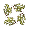
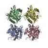
- Assembly
| Deposited unit | 
| |||||||||||||||||||||||||||||||||||||||||||||||||||||||||||||||||||||||||||||||||||||||||||||||||||||||||||||||||||||||||||||||||||||||||||||||||||||||||||||||||||||||||||||||||||||||||||||||||||||||||||||||||||||||||||||||||||||||||||||||||||||||||||||||||||||||||||||||||||||||||||||||||||||||||||||||||||||||||||
|---|---|---|---|---|---|---|---|---|---|---|---|---|---|---|---|---|---|---|---|---|---|---|---|---|---|---|---|---|---|---|---|---|---|---|---|---|---|---|---|---|---|---|---|---|---|---|---|---|---|---|---|---|---|---|---|---|---|---|---|---|---|---|---|---|---|---|---|---|---|---|---|---|---|---|---|---|---|---|---|---|---|---|---|---|---|---|---|---|---|---|---|---|---|---|---|---|---|---|---|---|---|---|---|---|---|---|---|---|---|---|---|---|---|---|---|---|---|---|---|---|---|---|---|---|---|---|---|---|---|---|---|---|---|---|---|---|---|---|---|---|---|---|---|---|---|---|---|---|---|---|---|---|---|---|---|---|---|---|---|---|---|---|---|---|---|---|---|---|---|---|---|---|---|---|---|---|---|---|---|---|---|---|---|---|---|---|---|---|---|---|---|---|---|---|---|---|---|---|---|---|---|---|---|---|---|---|---|---|---|---|---|---|---|---|---|---|---|---|---|---|---|---|---|---|---|---|---|---|---|---|---|---|---|---|---|---|---|---|---|---|---|---|---|---|---|---|---|---|---|---|---|---|---|---|---|---|---|---|---|---|---|---|---|---|---|---|---|---|---|---|---|---|---|---|---|---|---|---|---|---|---|---|---|---|---|---|---|---|---|---|---|---|---|---|---|---|---|---|---|---|---|---|---|---|---|---|---|---|---|---|---|---|---|---|---|---|
| 1 | 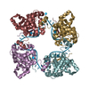
| |||||||||||||||||||||||||||||||||||||||||||||||||||||||||||||||||||||||||||||||||||||||||||||||||||||||||||||||||||||||||||||||||||||||||||||||||||||||||||||||||||||||||||||||||||||||||||||||||||||||||||||||||||||||||||||||||||||||||||||||||||||||||||||||||||||||||||||||||||||||||||||||||||||||||||||||||||||||||||
| 2 | 
| |||||||||||||||||||||||||||||||||||||||||||||||||||||||||||||||||||||||||||||||||||||||||||||||||||||||||||||||||||||||||||||||||||||||||||||||||||||||||||||||||||||||||||||||||||||||||||||||||||||||||||||||||||||||||||||||||||||||||||||||||||||||||||||||||||||||||||||||||||||||||||||||||||||||||||||||||||||||||||
| Unit cell |
| |||||||||||||||||||||||||||||||||||||||||||||||||||||||||||||||||||||||||||||||||||||||||||||||||||||||||||||||||||||||||||||||||||||||||||||||||||||||||||||||||||||||||||||||||||||||||||||||||||||||||||||||||||||||||||||||||||||||||||||||||||||||||||||||||||||||||||||||||||||||||||||||||||||||||||||||||||||||||||
| Noncrystallographic symmetry (NCS) | NCS domain:
NCS domain segments: Component-ID: _ / Refine code: _
|