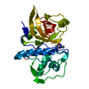
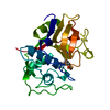
NEK9 subfamily protein kinase / nitric-oxide synthase inhibitor activity / negative regulation of DNA strand resection involved in replication fork processing / negative regulation of phosphorylation / intraciliary retrograde transport / Activation of BIM and translocation to mitochondria / Activation of NIMA Kinases NEK9, NEK6, NEK7 / motile cilium assembly / Nuclear Pore Complex (NPC) Disassembly / Intraflagellar transport ...NEK9 subfamily protein kinase / nitric-oxide synthase inhibitor activity / negative regulation of DNA strand resection involved in replication fork processing / negative regulation of phosphorylation / intraciliary retrograde transport / Activation of BIM and translocation to mitochondria / Activation of NIMA Kinases NEK9, NEK6, NEK7 / motile cilium assembly / Nuclear Pore Complex (NPC) Disassembly / Intraflagellar transport / positive regulation of intracellular transport / negative regulation of nitric oxide biosynthetic process / COPI-independent Golgi-to-ER retrograde traffic / cytoplasmic dynein complex / Macroautophagy / dynein intermediate chain binding / tertiary granule membrane / ficolin-1-rich granule membrane / spermatid development / protein kinase activator activity / COPI-mediated anterograde transport / Amplification of signal from unattached kinetochores via a MAD2 inhibitory signal / Loss of Nlp from mitotic centrosomes / Loss of proteins required for interphase microtubule organization from the centrosome / axon cytoplasm / Recruitment of mitotic centrosome proteins and complexes / MHC class II antigen presentation / Recruitment of NuMA to mitotic centrosomes / Anchoring of the basal body to the plasma membrane / substantia nigra development / Mitotic Prometaphase / HSP90 chaperone cycle for steroid hormone receptors (SHR) in the presence of ligand / EML4 and NUDC in mitotic spindle formation / regulation of mitotic cell cycle / enzyme inhibitor activity / ciliary tip / AURKA Activation by TPX2 / Resolution of Sister Chromatid Cohesion / positive regulation of insulin secretion involved in cellular response to glucose stimulus / RHO GTPases Activate Formins / kinetochore / HCMV Early Events / Aggrephagy / mitotic spindle / Separation of Sister Chromatids / Regulation of PLK1 Activity at G2/M Transition / mitotic cell cycle / site of double-strand break / scaffold protein binding / cytoskeleton / microtubule / non-specific serine/threonine protein kinase / cilium / cell division / protein serine kinase activity / protein serine/threonine kinase activity / apoptotic process / DNA damage response / Neutrophil degranulation / centrosome / protein kinase binding / protein-containing complex binding / enzyme binding / mitochondrion / ATP binding / membrane / metal ion binding / identical protein binding / nucleus / plasma membrane / cytoplasm / cytosol Similarity search - Function
Serine/threonine-protein kinase Nek9, catalytic domain / : / Protein Inhibitor Of Neuronal Nitric Oxide Synthase / Protein Inhibitor Of Neuronal Nitric Oxide Synthase; / : / RCC1-like domain / Regulator of chromosome condensation, RCC1 / Regulator of chromosome condensation (RCC1) repeat profile. / Dynein light chain, type 1/2, conserved site / Dynein light chain type 1 signature. ...Serine/threonine-protein kinase Nek9, catalytic domain / : / Protein Inhibitor Of Neuronal Nitric Oxide Synthase / Protein Inhibitor Of Neuronal Nitric Oxide Synthase; / : / RCC1-like domain / Regulator of chromosome condensation, RCC1 / Regulator of chromosome condensation (RCC1) repeat profile. / Dynein light chain, type 1/2, conserved site / Dynein light chain type 1 signature. / Dynein light chain, type 1/2 / Dynein light chain type 1 / Dynein light chain type 1 / Regulator of chromosome condensation 1/beta-lactamase-inhibitor protein II / Dynein light chain superfamily / Serine/threonine-protein kinase, active site / Serine/Threonine protein kinases active-site signature. / Protein kinase domain / Serine/Threonine protein kinases, catalytic domain / Protein kinase domain profile. / Protein kinase domain / Protein kinase-like domain superfamily / 2-Layer Sandwich / Alpha Beta Similarity search - Domain/homology
In the structure databanks used in Yorodumi, some data are registered as the other names, "COVID-19 virus" and "2019-nCoV". Here are the details of the virus and the list of structure data.
Jan 31, 2019. EMDB accession codes are about to change! (news from PDBe EMDB page)
EMDB accession codes are about to change! (news from PDBe EMDB page)
The allocation of 4 digits for EMDB accession codes will soon come to an end. Whilst these codes will remain in use, new EMDB accession codes will include an additional digit and will expand incrementally as the available range of codes is exhausted. The current 4-digit format prefixed with “EMD-” (i.e. EMD-XXXX) will advance to a 5-digit format (i.e. EMD-XXXXX), and so on. It is currently estimated that the 4-digit codes will be depleted around Spring 2019, at which point the 5-digit format will come into force.
The EM Navigator/Yorodumi systems omit the EMD- prefix.
Related info.:Q: What is EMD? / ID/Accession-code notation in Yorodumi/EM Navigator
Yorodumi is a browser for structure data from EMDB, PDB, SASBDB, etc.
This page is also the successor to EM Navigator detail page, and also detail information page/front-end page for Omokage search.
The word "yorodu" (or yorozu) is an old Japanese word meaning "ten thousand". "mi" (miru) is to see.
Related info.:EMDB / PDB / SASBDB / Comparison of 3 databanks / Yorodumi Search / Aug 31, 2016. New EM Navigator & Yorodumi / Yorodumi Papers / Jmol/JSmol / Function and homology information / Changes in new EM Navigator and Yorodumi
 Movie
Movie Controller


 Open data
 Basic information
 Components
 Keywords
 Function and homology information
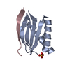
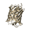
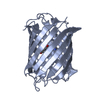
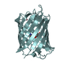
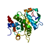
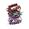
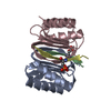
 HOMO SAPIENS (human)
 X-RAY DIFFRACTION /
  Authors
 Citation
 Structure visualization
 Downloads & links
 Other downloads


 PDBj
PDBj


 Assembly


 Mass: 18.015 Da / Num. of mol.: 19 / Source method: isolated from a natural source / Formula: H2O
 Sample preparation
 / Beamline: XALOC / Wavelength: 0.979494
  Processing