 Movie
Movie Controller


+ Open data

- Basic information
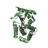
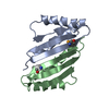
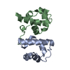
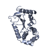
| Entry | Database: PDB / ID: 3zig | ||||||
|---|---|---|---|---|---|---|---|
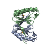
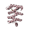
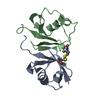
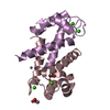
| Title | SepF-like protein from Pyrococcus furiosus | ||||||
 Components Components | SEPF-LIKE PROTEIN | ||||||
 Keywords Keywords | CELL CYCLE | ||||||
| Function / homology | Cell division protein SepF, archaea / SepF-like protein / Cell division protein SepF/SepF-related / Cell division protein SepF / SepF-like superfamily / Translation Initiation Factor IF3 / 2-Layer Sandwich / Alpha Beta / Cell division protein SepF Function and homology information Function and homology information | ||||||
| Biological species |   PYROCOCCUS FURIOSUS COM1 (archaea) PYROCOCCUS FURIOSUS COM1 (archaea) | ||||||
| Method |  X-RAY DIFFRACTION / SYNCHROTRON / MAD / Resolution: 2.5 Å X-RAY DIFFRACTION / SYNCHROTRON / MAD / Resolution: 2.5 Å | ||||||
 Authors Authors | Duman, R. / Ishikawa, S. / Celik, I. / Ogasawara, N. / Lowe, J. / Hamoen, L.W. | ||||||
 Citation Citation | Journal: Proc.Natl.Acad.Sci.USA / Year: 2013 Title: Structural and Genetic Analyses Reveal the Protein Sepf as a New Membrane Anchor for the Z Ring. Authors: Duman, R. / Ishikawa, S. / Celik, I. / Strahl, H. / Ogasawara, N. / Troc, P. / Lowe, J. / Hamoen, L.W. | ||||||
| History |
|
- Structure visualization
| Structure viewer | Molecule: MolmilJmol/JSmol |
|---|
- Downloads & links
-Download
| PDBx/mmCIF format | 3zig.cif.gz | 74.9 KB | Display | PDBx/mmCIF format |
|---|---|---|---|---|
| PDB format | pdb3zig.ent.gz | 57.7 KB | Display | PDB format |
| PDBx/mmJSON format | 3zig.json.gz | Tree view | PDBx/mmJSON format | |
| Others |  Other downloads Other downloads |
-Validation report
| Arichive directory | https://data.pdbj.org/pub/pdb/validation_reports/zi/3zigftp://data.pdbj.org/pub/pdb/validation_reports/zi/3zig | HTTPS FTP |
|---|
-Related structure data


-Links
 PDBj
PDBj- Assembly
| Deposited unit | 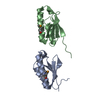
| |||||||||||||||||||||||||||||||||||||||||||||
|---|---|---|---|---|---|---|---|---|---|---|---|---|---|---|---|---|---|---|---|---|---|---|---|---|---|---|---|---|---|---|---|---|---|---|---|---|---|---|---|---|---|---|---|---|---|---|
| 1 | 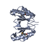
| |||||||||||||||||||||||||||||||||||||||||||||
| 2 | 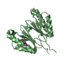
| |||||||||||||||||||||||||||||||||||||||||||||
| Unit cell |
| |||||||||||||||||||||||||||||||||||||||||||||
| Components on special symmetry positions |
| |||||||||||||||||||||||||||||||||||||||||||||
| Noncrystallographic symmetry (NCS) | NCS domain:
NCS domain segments:
NCS ensembles :
NCS oper:
|

-Components
| #1: Protein | Mass: 9496.246 Da / Num. of mol.: 2 / Fragment: RESIDUES 46-131 Source method: isolated from a genetically manipulated source Source: (gene. exp.) PYROCOCCUS FURIOSUS COM1 (archaea) / Plasmid: PHIS17 / Production host:  #2: Water | ChemComp-HOH / |  Mass: 18.015 Da / Num. of mol.: 29 / Source method: isolated from a natural source / Formula: H2O Mass: 18.015 Da / Num. of mol.: 29 / Source method: isolated from a natural source / Formula: H2OHas protein modification | Y | |
|---|
-Experimental details
-Experiment
| Experiment | Method: X-RAY DIFFRACTION / Number of used crystals: 1 |
|---|
- Sample preparation
| Crystal | Density Matthews: 2.21 Å3/Da / Density % sol: 44.38 % / Description: NONE |
|---|---|
| Crystal grow | Details: 0.2 M AMMONIUM SULFATE, 0.1 M HEPES PH 7.5, 25% W/V PEG 3350 |
-Data collection
| Diffraction | Mean temperature: 100 K |
|---|---|
| Diffraction source | Source: SYNCHROTRON / Site: ESRF  / Beamline: ID23-1 / Wavelength: 0.9793 / Beamline: ID23-1 / Wavelength: 0.9793 |
| Detector | Type: ADSC QUANTUM 315r / Detector: CCD / Date: Sep 5, 2010 |
| Radiation | Protocol: SINGLE WAVELENGTH / Monochromatic (M) / Laue (L): M / Scattering type: x-ray |
| Radiation wavelength | Wavelength: 0.9793 Å / Relative weight: 1 |
| Reflection | Resolution: 2.5→29.58 Å / Num. obs: 7036 / % possible obs: 99.9 % / Observed criterion σ(I): 2 / Redundancy: 26.2 % / Biso Wilson estimate: 39.32 Å2 / Rmerge(I) obs: 0.08 / Net I/σ(I): 31.1 |
| Reflection shell | Resolution: 2.5→2.64 Å / Redundancy: 25.8 % / Rmerge(I) obs: 0.23 / Mean I/σ(I) obs: 14.6 / % possible all: 100 |
- Processing
| Software |
| |||||||||||||||||||||||||||||||||||||||||||||||||||||||||||||||||||||||||||
|---|---|---|---|---|---|---|---|---|---|---|---|---|---|---|---|---|---|---|---|---|---|---|---|---|---|---|---|---|---|---|---|---|---|---|---|---|---|---|---|---|---|---|---|---|---|---|---|---|---|---|---|---|---|---|---|---|---|---|---|---|---|---|---|---|---|---|---|---|---|---|---|---|---|---|---|---|
| Refinement | Method to determine structure: MAD Starting model: NONE Resolution: 2.5→29.581 Å / SU ML: 0.4 / σ(F): 1.59 / Phase error: 27.52 / Stereochemistry target values: ML
| |||||||||||||||||||||||||||||||||||||||||||||||||||||||||||||||||||||||||||
| Solvent computation | Shrinkage radii: 0.83 Å / VDW probe radii: 1.1 Å / Solvent model: FLAT BULK SOLVENT MODEL / Bsol: 48.815 Å2 / ksol: 0.351 e/Å3 | |||||||||||||||||||||||||||||||||||||||||||||||||||||||||||||||||||||||||||
| Displacement parameters |
| |||||||||||||||||||||||||||||||||||||||||||||||||||||||||||||||||||||||||||
| Refinement step | Cycle: LAST / Resolution: 2.5→29.581 Å
| |||||||||||||||||||||||||||||||||||||||||||||||||||||||||||||||||||||||||||
| Refine LS restraints |
| |||||||||||||||||||||||||||||||||||||||||||||||||||||||||||||||||||||||||||
| Refine LS restraints NCS |
| |||||||||||||||||||||||||||||||||||||||||||||||||||||||||||||||||||||||||||
| LS refinement shell |
| |||||||||||||||||||||||||||||||||||||||||||||||||||||||||||||||||||||||||||
| Refinement TLS params. | Method: refined / Refine-ID: X-RAY DIFFRACTION
| |||||||||||||||||||||||||||||||||||||||||||||||||||||||||||||||||||||||||||
| Refinement TLS group |
|
