 Movie
Movie Controller

[English] 日本語
 Yorodumi
- PDB-3zf4: Phage dUTPases control transfer of virulence genes by a proto- on... -
+ Open data

- Basic information
| Entry | Database: PDB / ID: 3zf4 | ||||||
|---|---|---|---|---|---|---|---|
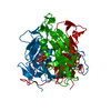
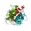
| Title | Phage dUTPases control transfer of virulence genes by a proto- oncogenic G protein-like mechanism. (Staphylococcus bacteriophage 80alpha dUTPase Y81A mutant with dUpNHpp). | ||||||
 Components Components | DUTPASE | ||||||
 Keywords Keywords | HYDROLASE / PATHOGENICITY ISLAND / SAPI INDUCTION / GENE TRANSFER / MOONLIGHTING PROTEINS / G-PROTEIN / P-LOOP | ||||||
| Function / homology |  Function and homology information Function and homology informationdUTP catabolic process / dUMP biosynthetic process / dUTP diphosphatase / dUTP diphosphatase activity / magnesium ion binding Similarity search - Function | ||||||
| Biological species |  STAPHYLOCOCCUS PHAGE 80ALPHA (virus) STAPHYLOCOCCUS PHAGE 80ALPHA (virus) | ||||||
| Method |  X-RAY DIFFRACTION / SYNCHROTRON / MOLECULAR REPLACEMENT / Resolution: 3.1 Å X-RAY DIFFRACTION / SYNCHROTRON / MOLECULAR REPLACEMENT / Resolution: 3.1 Å | ||||||
 Authors Authors | Tormo-Mas, M.A. / Donderis, J. / Garcia-Caballer, M. / Alt, A. / Mir-Sanchis, I. / Marina, A. / Penades, J.R. | ||||||
 Citation Citation | Journal: Mol.Cell / Year: 2013 Title: Phage Dutpases Control Transfer of Virulence Genes by a Proto-Oncogenic G Protein-Like Mechanism. Authors: Tormo-Mas, M.A. / Donderis, J. / Garcia-Caballer, M. / Alt, A. / Mir-Sanchis, I. / Marina, A. / Penades, J.R. #1: Journal: Nature / Year: 2010 Title: Moonlighting Bacteriophage Proteins Derepress Staphylococcal Pathogenicity Islands. Authors: Tormo-Mas, M.A. / Mir, I. / Shrestha, A. / Tallent, S.M. / Campoy, S. / Lasa, I. / Barbe, J. / Novick, R.P. / Christie, G.E. / Penades, J.R. | ||||||
| History |
|
- Structure visualization
| Structure viewer | Molecule: MolmilJmol/JSmol |
|---|
- Downloads & links
-Download
| PDBx/mmCIF format | 3zf4.cif.gz | 50.1 KB | Display | PDBx/mmCIF format |
|---|---|---|---|---|
| PDB format | pdb3zf4.ent.gz | 33.6 KB | Display | PDB format |
| PDBx/mmJSON format | 3zf4.json.gz | Tree view | PDBx/mmJSON format | |
| Others |  Other downloads Other downloads |
-Validation report
| Arichive directory | https://data.pdbj.org/pub/pdb/validation_reports/zf/3zf4ftp://data.pdbj.org/pub/pdb/validation_reports/zf/3zf4 | HTTPS FTP |
|---|
-Related structure data
| Related structure data |  3zezSC  3zf0C  3zf1C  3zf2C  3zf3C 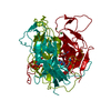 3zf5C 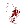 3zf6C S: Starting model for refinement C: citing same article ( |
|---|---|
| Similar structure data |


-Links
 PDBj
PDBj
- Assembly
| Deposited unit | 
| |||||||||
|---|---|---|---|---|---|---|---|---|---|---|
| 1 | 
| |||||||||
| Unit cell |
| |||||||||
| Components on special symmetry positions |
|
-Components
| #1: Protein | Mass: 22575.529 Da / Num. of mol.: 1 / Mutation: YES Source method: isolated from a genetically manipulated source Details: STRUCTURE IN PRESENCE OF DUPNHPP AND MAGNESIUM / Source: (gene. exp.) STAPHYLOCOCCUS PHAGE 80ALPHA (virus) / Production host:  | ||
|---|---|---|---|
| #2: Chemical | ChemComp-DUP /   Mass: 467.157 Da / Num. of mol.: 1 / Source method: obtained synthetically / Formula: C9H16N3O13P3 Mass: 467.157 Da / Num. of mol.: 1 / Source method: obtained synthetically / Formula: C9H16N3O13P3 | ||
| #3: Chemical | ChemComp-MG /   Mass: 24.305 Da / Num. of mol.: 1 / Source method: obtained synthetically / Formula: Mg Mass: 24.305 Da / Num. of mol.: 1 / Source method: obtained synthetically / Formula: Mg | ||
| #4: Chemical |   Mass: 58.693 Da / Num. of mol.: 2 / Source method: obtained synthetically / Formula: Ni Mass: 58.693 Da / Num. of mol.: 2 / Source method: obtained synthetically / Formula: Ni#5: Water | ChemComp-HOH / |  Mass: 18.015 Da / Num. of mol.: 12 / Source method: isolated from a natural source / Formula: H2O Mass: 18.015 Da / Num. of mol.: 12 / Source method: isolated from a natural source / Formula: H2O |
-Experimental details
-Experiment
| Experiment | Method: X-RAY DIFFRACTION |
|---|
- Sample preparation
| Crystal | Density Matthews: 2.44 Å3/Da / Density % sol: 49.6 % / Description: NONE |
|---|
-Data collection
| Diffraction | Mean temperature: 100 K |
|---|---|
| Diffraction source | Source: SYNCHROTRON / Site: Diamond  / Beamline: I04-1 / Wavelength: 0.9795 / Beamline: I04-1 / Wavelength: 0.9795 |
| Radiation | Protocol: SINGLE WAVELENGTH / Monochromatic (M) / Laue (L): M / Scattering type: x-ray |
| Radiation wavelength | Wavelength: 0.9795 Å / Relative weight: 1 |
| Reflection | Resolution: 3.1→61.9 Å / Num. obs: 4224 / % possible obs: 99.7 % / Observed criterion σ(I): 2 / Redundancy: 5.1 % / Rmerge(I) obs: 0.06 / Net I/σ(I): 8.2 |
| Reflection shell | Resolution: 3.1→3.27 Å / Redundancy: 5.3 % / Rmerge(I) obs: 0.33 / Mean I/σ(I) obs: 2.4 / % possible all: 100 |
- Processing
| Software |
| ||||||||||||||||||||||||||||||||||||||||||||||||||||||||||||||||||||||||||||||||||||||||||||||||||||||||||||||||||||||||||||||||||||||||||||||||||||||||||||||||||||||||||||||||||||||
|---|---|---|---|---|---|---|---|---|---|---|---|---|---|---|---|---|---|---|---|---|---|---|---|---|---|---|---|---|---|---|---|---|---|---|---|---|---|---|---|---|---|---|---|---|---|---|---|---|---|---|---|---|---|---|---|---|---|---|---|---|---|---|---|---|---|---|---|---|---|---|---|---|---|---|---|---|---|---|---|---|---|---|---|---|---|---|---|---|---|---|---|---|---|---|---|---|---|---|---|---|---|---|---|---|---|---|---|---|---|---|---|---|---|---|---|---|---|---|---|---|---|---|---|---|---|---|---|---|---|---|---|---|---|---|---|---|---|---|---|---|---|---|---|---|---|---|---|---|---|---|---|---|---|---|---|---|---|---|---|---|---|---|---|---|---|---|---|---|---|---|---|---|---|---|---|---|---|---|---|---|---|---|---|
| Refinement | Method to determine structure: MOLECULAR REPLACEMENT Starting model: PDB ENTRY 3ZEZ Resolution: 3.1→61.85 Å / Cor.coef. Fo:Fc: 0.92 / Cor.coef. Fo:Fc free: 0.89 / SU B: 24.204 / SU ML: 0.424 / Cross valid method: THROUGHOUT / ESU R Free: 0.116 / Stereochemistry target values: MAXIMUM LIKELIHOOD Details: HYDROGENS HAVE BEEN ADDED IN THE RIDING POSITIONS. INITIAL MET IS NOT TRACED. RESIDUES K143 AND V170 ARE NOT TRACED BECAUSE OF LACK OF ELECTRON DENSITY. RESIUDES E159, , R160, K163, G169 ARE ...Details: HYDROGENS HAVE BEEN ADDED IN THE RIDING POSITIONS. INITIAL MET IS NOT TRACED. RESIDUES K143 AND V170 ARE NOT TRACED BECAUSE OF LACK OF ELECTRON DENSITY. RESIUDES E159, , R160, K163, G169 ARE TRACED AS A. RESIDUES S156-S158 ARE DISORDERED.
| ||||||||||||||||||||||||||||||||||||||||||||||||||||||||||||||||||||||||||||||||||||||||||||||||||||||||||||||||||||||||||||||||||||||||||||||||||||||||||||||||||||||||||||||||||||||
| Solvent computation | Ion probe radii: 0.8 Å / Shrinkage radii: 0.8 Å / VDW probe radii: 1.2 Å / Solvent model: MASK | ||||||||||||||||||||||||||||||||||||||||||||||||||||||||||||||||||||||||||||||||||||||||||||||||||||||||||||||||||||||||||||||||||||||||||||||||||||||||||||||||||||||||||||||||||||||
| Displacement parameters | Biso mean: 105.915 Å2
| ||||||||||||||||||||||||||||||||||||||||||||||||||||||||||||||||||||||||||||||||||||||||||||||||||||||||||||||||||||||||||||||||||||||||||||||||||||||||||||||||||||||||||||||||||||||
| Refinement step | Cycle: LAST / Resolution: 3.1→61.85 Å
| ||||||||||||||||||||||||||||||||||||||||||||||||||||||||||||||||||||||||||||||||||||||||||||||||||||||||||||||||||||||||||||||||||||||||||||||||||||||||||||||||||||||||||||||||||||||
| Refine LS restraints |
|