[English] 日本語
Yorodumi
- PDB-5nyz: Twist and induce: Dissecting the link between the enzymatic activ... -

+
Open data


ID or keywords:

Loading...

-
Basic information

Entry
Database: PDB / ID: 5nyz
Title
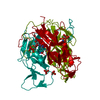
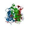
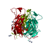
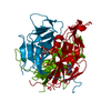
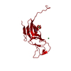
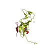
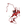
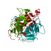
Twist and induce: Dissecting the link between the enzymatic activity and the SaPI inducing capacity of the phage 80 dUTPase. D95E mutant from dUTPase 80alpha phage.
ComponentsDUTPaseDUTP diphosphatase
KeywordsHYDROLASE / dUTPase / 80 phage / S.aureus
Function / homology
Function and homology information


dUTP catabolic process / dUMP biosynthetic process / dUTP diphosphatase / dUTP diphosphatase activity / magnesium ion binding
Similarity search - Function
Deoxyuridine triphosphate nucleotidohydrolase / Deoxyuridine triphosphatase (dUTPase) / Deoxyuridine 5'-Triphosphate Nucleotidohydrolase; Chain A / dUTPase-like / dUTPase / dUTPase, trimeric / dUTPase-like superfamily / Distorted Sandwich / Mainly Beta
Similarity search - Domain/homology
2'-DEOXYURIDINE 5'-ALPHA,BETA-IMIDO-TRIPHOSPHATE / dUTP diphosphatase
Similarity search - Component
Biological speciesStaphylococcus phage 80alpha (virus)
MethodX-RAY DIFFRACTION / SYNCHROTRON / MOLECULAR REPLACEMENT / Resolution: 2.5 Å
AuthorsAlite, C. / Humphrey, S. / Donderis, J. / Maiques, E. / Ciges-Tomas, J.R. / Penades, J.R. / Marina, A.
Funding support Spain, 2items
OrganizationGrant numberCountry
Spanish Ministry of Economy and CompetitivenessBES-2014-068617 Spain
Spanish Ministry of Economy and CompetitivenessBIO2013-42619-P Spain
CitationJournal: Sci Rep / Year: 2017
Title: Dissecting the link between the enzymatic activity and the SaPI inducing capacity of the phage 80 alpha dUTPase.
Authors: Alite, C. / Humphrey, S. / Donderis, J. / Maiques, E. / Ciges-Tomas, J.R. / Penades, J.R. / Marina, A.
History
DepositionMay 12, 2017Deposition site: PDBE / Processing site: PDBE
Revision 1.0Sep 20, 2017Provider: repository / Type: Initial release
Revision 1.1Jan 17, 2024Group: Data collection / Database references ...Data collection / Database references / Derived calculations / Refinement description
Category: chem_comp_atom / chem_comp_bond ...chem_comp_atom / chem_comp_bond / database_2 / pdbx_initial_refinement_model / pdbx_struct_conn_angle / struct_conn
Item: _database_2.pdbx_DOI / _database_2.pdbx_database_accession ..._database_2.pdbx_DOI / _database_2.pdbx_database_accession / _pdbx_struct_conn_angle.ptnr1_auth_comp_id / _pdbx_struct_conn_angle.ptnr1_auth_seq_id / _pdbx_struct_conn_angle.ptnr1_label_asym_id / _pdbx_struct_conn_angle.ptnr1_label_atom_id / _pdbx_struct_conn_angle.ptnr1_label_comp_id / _pdbx_struct_conn_angle.ptnr1_label_seq_id / _pdbx_struct_conn_angle.ptnr2_auth_seq_id / _pdbx_struct_conn_angle.ptnr2_label_asym_id / _pdbx_struct_conn_angle.ptnr2_symmetry / _pdbx_struct_conn_angle.ptnr3_auth_comp_id / _pdbx_struct_conn_angle.ptnr3_auth_seq_id / _pdbx_struct_conn_angle.ptnr3_label_asym_id / _pdbx_struct_conn_angle.ptnr3_label_atom_id / _pdbx_struct_conn_angle.ptnr3_label_comp_id / _pdbx_struct_conn_angle.ptnr3_label_seq_id / _pdbx_struct_conn_angle.ptnr3_symmetry / _pdbx_struct_conn_angle.value / _struct_conn.pdbx_dist_value / _struct_conn.ptnr1_auth_comp_id / _struct_conn.ptnr1_auth_seq_id / _struct_conn.ptnr1_label_asym_id / _struct_conn.ptnr1_label_atom_id / _struct_conn.ptnr1_label_comp_id / _struct_conn.ptnr1_label_seq_id / _struct_conn.ptnr2_auth_comp_id / _struct_conn.ptnr2_auth_seq_id / _struct_conn.ptnr2_label_asym_id / _struct_conn.ptnr2_label_atom_id / _struct_conn.ptnr2_label_comp_id / _struct_conn.ptnr2_symmetry

-
Structure visualization

Structure viewerMolecule:
MolmilJmol/JSmol

Downloads & links

-
Assembly

Deposited unit
A: DUTPase
hetero molecules


Theoretical massNumber of molelcules
Total (without water)19,6444
Polymers19,1291
Non-polymers5163
Water25214
1
A: DUTPase
hetero molecules

A: DUTPase
hetero molecules

A: DUTPase
hetero molecules


Theoretical massNumber of molelcules
Total (without water)58,93312
Polymers57,3863
Non-polymers1,5479
Water543
TypeNameSymmetry operationNumber
identity operation1_555x,y,z1
crystal symmetry operation7_555-z+1/2,-x,y+1/21
crystal symmetry operation10_545-y,z-1/2,-x+1/21
Buried area16160 Å2
ΔGint-89 kcal/mol
Surface area20870 Å2
MethodPISA
Unit cell
Length a, b, c (Å)87.270, 87.270, 87.270
Angle α, β, γ (deg.)90.00, 90.00, 90.00
Int Tables number198
Space group name H-MP213
Components on special symmetry positions
IDModelComponents
11A-312-

HOH

-
Components

#1: Protein DUTPase / DUTP diphosphatase


Mass: 19128.695 Da / Num. of mol.: 1 / Mutation: D95E
Source method: isolated from a genetically manipulated source
Details: Ordered C-terminal is placed over the nucleotide on active center. Magnesium atom is coordinated by E95 residue.
Source: (gene. exp.) Staphylococcus phage 80alpha (virus) / Plasmid: pet28a / Production host: Escherichia coli BL21(DE3) (bacteria) / References: UniProt: A4ZF98
#2: Chemical ChemComp-DUP / 2'-DEOXYURIDINE 5'-ALPHA,BETA-IMIDO-TRIPHOSPHATE


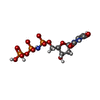
Mass: 467.157 Da / Num. of mol.: 1 / Source method: obtained synthetically / Formula: C9H16N3O13P3
#3: Chemical ChemComp-MG / MAGNESIUM ION


Mass: 24.305 Da / Num. of mol.: 2 / Source method: obtained synthetically / Formula: Mg
#4: Water ChemComp-HOH / water / Water


Mass: 18.015 Da / Num. of mol.: 14 / Source method: isolated from a natural source / Formula: H2O

-
Experimental details

-
Experiment

ExperimentMethod: X-RAY DIFFRACTION / Number of used crystals: 1

-
Sample preparation

CrystalDensity Matthews: 2.41 Å3/Da / Density % sol: 48.96 %
Crystal growTemperature: 296 K / Method: vapor diffusion, sitting drop / pH: 7.5 / Details: 20% Ethanol; 10% Glicerol

-
Data collection

DiffractionMean temperature: 100 K
Diffraction sourceSource: SYNCHROTRON / Site: ALBA / Beamline: XALOC / Wavelength: 0.97921 Å
DetectorType: DECTRIS PILATUS3 6M / Detector: PIXEL / Date: Apr 15, 2016
RadiationProtocol: SINGLE WAVELENGTH / Monochromatic (M) / Laue (L): M / Scattering type: x-ray
Radiation wavelengthWavelength: 0.97921 Å / Relative weight: 1
ReflectionResolution: 2.5→87.27 Å / Num. obs: 7924 / % possible obs: 100 % / Redundancy: 18 % / CC1/2: 0.898 / Rpim(I) all: 0.038 / Net I/σ(I): 15.4
Reflection shellResolution: 2.5→2.6 Å / Redundancy: 18.1 % / Mean I/σ(I) obs: 2 / Num. unique obs: 882 / CC1/2: 0.658 / Rpim(I) all: 0.573 / % possible all: 100

-
Processing

Software
NameVersionClassification
REFMAC5.8.0158refinement
iMOSFLMdata reduction
Aimlessdata scaling
PHASERphasing
RefinementMethod to determine structure: MOLECULAR REPLACEMENT
Starting model: 3ZEZ
Resolution: 2.5→61.71 Å / Cor.coef. Fo:Fc: 0.938 / Cor.coef. Fo:Fc free: 0.894 / SU B: 11.95 / SU ML: 0.265 / Cross valid method: THROUGHOUT / ESU R: 0.452 / ESU R Free: 0.301 / Details: HYDROGENS HAVE BEEN ADDED IN THE RIDING POSITIONS
RfactorNum. reflection% reflectionSelection details
Rfree0.28055 374 4.7 %RANDOM
Rwork0.22569 ---
obs0.22853 7533 100 %-
Solvent computationIon probe radii: 0.8 Å / Shrinkage radii: 0.8 Å / VDW probe radii: 1.2 Å
Displacement parametersBiso mean: 60.162 Å2
Baniso -1Baniso -2Baniso -3
1-0 Å20 Å20 Å2
2--0 Å20 Å2
3---0 Å2
Refinement stepCycle: 1 / Resolution: 2.5→61.71 Å
ProteinNucleic acidLigandSolventTotal
Num. atoms1299 0 30 14 1343
Refine LS restraints
Refine-IDTypeDev idealDev ideal targetNumber
X-RAY DIFFRACTIONr_bond_refined_d0.0060.0191348
X-RAY DIFFRACTIONr_bond_other_d0.0010.021239
X-RAY DIFFRACTIONr_angle_refined_deg1.141.9851826
X-RAY DIFFRACTIONr_angle_other_deg0.82332874
X-RAY DIFFRACTIONr_dihedral_angle_1_deg5.8735165
X-RAY DIFFRACTIONr_dihedral_angle_2_deg32.67924.75461
X-RAY DIFFRACTIONr_dihedral_angle_3_deg13.02615236
X-RAY DIFFRACTIONr_dihedral_angle_4_deg16.01158
X-RAY DIFFRACTIONr_chiral_restr0.0660.2207
X-RAY DIFFRACTIONr_gen_planes_refined0.0030.021475
X-RAY DIFFRACTIONr_gen_planes_other0.0010.02257
X-RAY DIFFRACTIONr_nbd_refined
X-RAY DIFFRACTIONr_nbd_other
X-RAY DIFFRACTIONr_nbtor_refined
X-RAY DIFFRACTIONr_nbtor_other
X-RAY DIFFRACTIONr_xyhbond_nbd_refined
X-RAY DIFFRACTIONr_xyhbond_nbd_other
X-RAY DIFFRACTIONr_metal_ion_refined
X-RAY DIFFRACTIONr_metal_ion_other
X-RAY DIFFRACTIONr_symmetry_vdw_refined
X-RAY DIFFRACTIONr_symmetry_vdw_other
X-RAY DIFFRACTIONr_symmetry_hbond_refined
X-RAY DIFFRACTIONr_symmetry_hbond_other
X-RAY DIFFRACTIONr_symmetry_metal_ion_refined
X-RAY DIFFRACTIONr_symmetry_metal_ion_other
X-RAY DIFFRACTIONr_mcbond_it1.8096.14666
X-RAY DIFFRACTIONr_mcbond_other1.8086.135665
X-RAY DIFFRACTIONr_mcangle_it3.1539.18829
X-RAY DIFFRACTIONr_mcangle_other3.1519.187830
X-RAY DIFFRACTIONr_scbond_it1.6296.435682
X-RAY DIFFRACTIONr_scbond_other1.6316.433681
X-RAY DIFFRACTIONr_scangle_it
X-RAY DIFFRACTIONr_scangle_other2.8799.568998
X-RAY DIFFRACTIONr_long_range_B_refined5.46168.551316
X-RAY DIFFRACTIONr_long_range_B_other5.3368.5281316
X-RAY DIFFRACTIONr_rigid_bond_restr
X-RAY DIFFRACTIONr_sphericity_free
X-RAY DIFFRACTIONr_sphericity_bonded
LS refinement shellResolution: 2.501→2.565 Å / Total num. of bins used: 20
RfactorNum. reflection% reflection
Rfree0.384 20 -
Rwork0.316 559 -
obs--100 %

+
About Yorodumi

-
News

-
Feb 9, 2022. New format data for meta-information of EMDB entries

New format data for meta-information of EMDB entries

  • Version 3 of the EMDB header file is now the official format.
  • The previous official version 1.9 will be removed from the archive.

Related info.:EMDB header

External links:wwPDB to switch to version 3 of the EMDB data model

-
Aug 12, 2020. Covid-19 info

Covid-19 info

URL: https://pdbj.org/emnavi/covid19.php

New page: Covid-19 featured information page in EM Navigator.

Related info.:Covid-19 info / Mar 5, 2020. Novel coronavirus structure data

+
Mar 5, 2020. Novel coronavirus structure data

Novel coronavirus structure data

Related info.:Yorodumi Speices / Aug 12, 2020. Covid-19 info

External links:COVID-19 featured content - PDBj / Molecule of the Month (242):Coronavirus Proteases

+
Jan 31, 2019. EMDB accession codes are about to change! (news from PDBe EMDB page)

EMDB accession codes are about to change! (news from PDBe EMDB page)

  • The allocation of 4 digits for EMDB accession codes will soon come to an end. Whilst these codes will remain in use, new EMDB accession codes will include an additional digit and will expand incrementally as the available range of codes is exhausted. The current 4-digit format prefixed with “EMD-” (i.e. EMD-XXXX) will advance to a 5-digit format (i.e. EMD-XXXXX), and so on. It is currently estimated that the 4-digit codes will be depleted around Spring 2019, at which point the 5-digit format will come into force.
  • The EM Navigator/Yorodumi systems omit the EMD- prefix.

Related info.:Q: What is EMD? / ID/Accession-code notation in Yorodumi/EM Navigator

External links:EMDB Accession Codes are Changing Soon! / Contact to PDBj

+
Jul 12, 2017. Major update of PDB

Major update of PDB

  • wwPDB released updated PDB data conforming to the new PDBx/mmCIF dictionary.
  • This is a major update changing the version number from 4 to 5, and with Remediation, in which all the entries are updated.
  • In this update, many items about electron microscopy experimental information are reorganized (e.g. em_software).
  • Now, EM Navigator and Yorodumi are based on the updated data.

External links:wwPDB Remediation / Enriched Model Files Conforming to OneDep Data Standards Now Available in the PDB FTP Archive

-
Yorodumi

Thousand views of thousand structures

  • Yorodumi is a browser for structure data from EMDB, PDB, SASBDB, etc.
  • This page is also the successor to EM Navigator detail page, and also detail information page/front-end page for Omokage search.
  • The word "yorodu" (or yorozu) is an old Japanese word meaning "ten thousand". "mi" (miru) is to see.

Related info.:EMDB / PDB / SASBDB / Comparison of 3 databanks / Yorodumi Search / Aug 31, 2016. New EM Navigator & Yorodumi / Yorodumi Papers / Jmol/JSmol / Function and homology information / Changes in new EM Navigator and Yorodumi

Read more