 Movie
Movie Controller

[English] 日本語
 Yorodumi
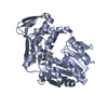
- PDB-3zda: Structure of E. coli ExoIX in complex with a fragment of the Flap... -
+ Open data

- Basic information
| Entry | Database: PDB / ID: 3zda | ||||||
|---|---|---|---|---|---|---|---|
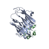
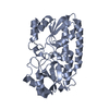
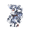
| Title | Structure of E. coli ExoIX in complex with a fragment of the Flap1 DNA oligonucleotide, potassium and magnesium | ||||||
 Components Components |
| ||||||
 Keywords Keywords | HYDROLASE/DNA / HYDROLASE-DNA COMPLEX / FLAP ENDONUCLEASE / DNA BINDING | ||||||
| Function / homology |  Function and homology information Function and homology informationDNA replication, Okazaki fragment processing / 5'-flap endonuclease activity / 5'-3' exonuclease activity / potassium ion binding / Hydrolases; Acting on ester bonds / magnesium ion binding / DNA binding Similarity search - Function | ||||||
| Biological species |  SYNTHETIC CONSTRUCT (others) | ||||||
| Method |  X-RAY DIFFRACTION / SYNCHROTRON / MOLECULAR REPLACEMENT / Resolution: 1.5 Å X-RAY DIFFRACTION / SYNCHROTRON / MOLECULAR REPLACEMENT / Resolution: 1.5 Å | ||||||
 Authors Authors | Hemsworth, G.R. / Anstey-Gilbert, C.S. / Flemming, C.S. / Hodskinson, M.R.G. / Zhang, J. / Sedelnikova, S.E. / Stillman, T.J. / Sayers, J.R. / Artymiuk, P.J. | ||||||
 Citation Citation | Journal: Nucleic Acids Res. / Year: 2013 Title: The Structure of E. Coli Exoix - Implications for DNA Binding and Catalysis in Flap Endonucleases Authors: Anstey-Gilbert, C.S. / Hemsworth, G.R. / Flemming, C.S. / Hodskinson, M.R.G. / Zhang, J. / Sedelnikova, S.E. / Stillman, T.J. / Sayers, J.R. / Artymiuk, P.J. | ||||||
| History |
|
- Structure visualization
| Structure viewer | Molecule: MolmilJmol/JSmol |
|---|
- Downloads & links
-Download
| PDBx/mmCIF format | 3zda.cif.gz | 78.8 KB | Display | PDBx/mmCIF format |
|---|---|---|---|---|
| PDB format | pdb3zda.ent.gz | 54.7 KB | Display | PDB format |
| PDBx/mmJSON format | 3zda.json.gz | Tree view | PDBx/mmJSON format | |
| Others |  Other downloads Other downloads |
-Validation report
| Summary document | 3zda_validation.pdf.gz | 464.3 KB | Display | wwPDB validaton report |
|---|---|---|---|---|
| Full document | 3zda_full_validation.pdf.gz | 466.5 KB | Display | |
| Data in XML | 3zda_validation.xml.gz | 14.6 KB | Display | |
| Data in CIF | 3zda_validation.cif.gz | 21.5 KB | Display | |
| Arichive directory | https://data.pdbj.org/pub/pdb/validation_reports/zd/3zdaftp://data.pdbj.org/pub/pdb/validation_reports/zd/3zda | HTTPS FTP |
-Related structure data
| Related structure data |  3zd8C  3zd9SC  3zdbC  3zdcC  3zddC  3zdeC C: citing same article ( S: Starting model for refinement |
|---|---|
| Similar structure data |


-Links
 PDBj
PDBj

- Assembly
| Deposited unit | 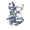
| ||||||||
|---|---|---|---|---|---|---|---|---|---|
| 1 |
| ||||||||
| Unit cell |
|
-Components
-Protein , 1 types, 1 molecules A
| #1: Protein | Mass: 28203.158 Da / Num. of mol.: 1 Source method: isolated from a genetically manipulated source Source: (gene. exp.) References: UniProt: Q8X6R9, Hydrolases; Acting on ester bonds |
|---|
-DNA chain , 2 types, 2 molecules BC
| #2: DNA chain | Mass: 1191.818 Da / Num. of mol.: 1 / Source method: obtained synthetically / Source: (synth.) SYNTHETIC CONSTRUCT (others) |
|---|---|
| #3: DNA chain | Mass: 1818.231 Da / Num. of mol.: 1 / Source method: obtained synthetically / Source: (synth.) SYNTHETIC CONSTRUCT (others) |
-Non-polymers , 5 types, 247 molecules 


| #4: Chemical | ChemComp-K /  Mass: 39.098 Da / Num. of mol.: 1 / Source method: obtained synthetically / Formula: K Mass: 39.098 Da / Num. of mol.: 1 / Source method: obtained synthetically / Formula: K |
|---|---|
| #5: Chemical | ChemComp-MG /  Mass: 24.305 Da / Num. of mol.: 1 / Source method: obtained synthetically / Formula: Mg Mass: 24.305 Da / Num. of mol.: 1 / Source method: obtained synthetically / Formula: Mg |
| #6: Chemical | ChemComp-PIV /  Type: L-peptide linking / Mass: 102.132 Da / Num. of mol.: 1 / Source method: obtained synthetically / Formula: C5H10O2 Type: L-peptide linking / Mass: 102.132 Da / Num. of mol.: 1 / Source method: obtained synthetically / Formula: C5H10O2 |
| #7: Chemical | ChemComp-PO4 /  Mass: 94.971 Da / Num. of mol.: 1 / Source method: obtained synthetically / Formula: PO4 Mass: 94.971 Da / Num. of mol.: 1 / Source method: obtained synthetically / Formula: PO4 |
| #8: Water | ChemComp-HOH / Mass: 18.015 Da / Num. of mol.: 243 / Source method: isolated from a natural source / Formula: H2O |
-Details
| Sequence details | FRAGMENT OF LARGER DNA BOUND TO PROTEIN. SEQUENCE IS ACTUALLY UNKNOWN, CRYSTAL LIKELY HAS MIXED ...FRAGMENT OF LARGER DNA BOUND TO PROTEIN. SEQUENCE IS ACTUALLY UNKNOWN, CRYSTAL LIKELY HAS MIXED DNAS BOUND HERE. |
|---|
-Experimental details
-Experiment
| Experiment | Method: X-RAY DIFFRACTION / Number of used crystals: 1 |
|---|
- Sample preparation
| Crystal | Density Matthews: 2.32 Å3/Da / Density % sol: 47 % / Description: NONE |
|---|---|
| Crystal grow | pH: 6 Details: 0.2 M MAGNESIUM ACETATE, 15% (W/V) PEG-3350, pH 6.0 |
-Data collection
| Diffraction | Mean temperature: 100 K |
|---|---|
| Diffraction source | Source: SYNCHROTRON / Site: Diamond  / Beamline: I03 / Wavelength: 0.976 / Beamline: I03 / Wavelength: 0.976 |
| Detector | Type: ADSC CCD / Detector: CCD / Date: Dec 16, 2008 / Details: MIRRORS |
| Radiation | Monochromator: DOUBLE CRYSTAL / Protocol: SINGLE WAVELENGTH / Monochromatic (M) / Laue (L): M / Scattering type: x-ray |
| Radiation wavelength | Wavelength: 0.976 Å / Relative weight: 1 |
| Reflection | Resolution: 1.5→51.64 Å / Num. obs: 57905 / % possible obs: 99.9 % / Observed criterion σ(I): 6 / Redundancy: 9.9 % / Rmerge(I) obs: 0.1 / Net I/σ(I): 22.5 |
| Reflection shell | Resolution: 1.5→1.58 Å / Redundancy: 7 % / Rmerge(I) obs: 0.48 / Mean I/σ(I) obs: 3.4 / % possible all: 99.8 |
- Processing
| Software |
| ||||||||||||||||||||||||||||||||||||||||||||||||||||||||||||||||||||||||||||||||||||||||||||||||||||||||||||||||||||||||||||||||||||||||||||||||||||||||||||||||||||||||||||||||||||||
|---|---|---|---|---|---|---|---|---|---|---|---|---|---|---|---|---|---|---|---|---|---|---|---|---|---|---|---|---|---|---|---|---|---|---|---|---|---|---|---|---|---|---|---|---|---|---|---|---|---|---|---|---|---|---|---|---|---|---|---|---|---|---|---|---|---|---|---|---|---|---|---|---|---|---|---|---|---|---|---|---|---|---|---|---|---|---|---|---|---|---|---|---|---|---|---|---|---|---|---|---|---|---|---|---|---|---|---|---|---|---|---|---|---|---|---|---|---|---|---|---|---|---|---|---|---|---|---|---|---|---|---|---|---|---|---|---|---|---|---|---|---|---|---|---|---|---|---|---|---|---|---|---|---|---|---|---|---|---|---|---|---|---|---|---|---|---|---|---|---|---|---|---|---|---|---|---|---|---|---|---|---|---|---|
| Refinement | Method to determine structure: MOLECULAR REPLACEMENT Starting model: PDB ENTRY 3ZD9 Resolution: 1.5→51.64 Å / Cor.coef. Fo:Fc: 0.954 / Cor.coef. Fo:Fc free: 0.941 / SU B: 1.333 / SU ML: 0.051 / Cross valid method: THROUGHOUT / ESU R: 0.074 / ESU R Free: 0.075 / Stereochemistry target values: MAXIMUM LIKELIHOOD / Details: HYDROGENS HAVE BEEN ADDED IN THE RIDING POSITIONS.
| ||||||||||||||||||||||||||||||||||||||||||||||||||||||||||||||||||||||||||||||||||||||||||||||||||||||||||||||||||||||||||||||||||||||||||||||||||||||||||||||||||||||||||||||||||||||
| Solvent computation | Ion probe radii: 0.8 Å / Shrinkage radii: 0.8 Å / VDW probe radii: 1.4 Å / Solvent model: MASK | ||||||||||||||||||||||||||||||||||||||||||||||||||||||||||||||||||||||||||||||||||||||||||||||||||||||||||||||||||||||||||||||||||||||||||||||||||||||||||||||||||||||||||||||||||||||
| Displacement parameters | Biso mean: 25.205 Å2
| ||||||||||||||||||||||||||||||||||||||||||||||||||||||||||||||||||||||||||||||||||||||||||||||||||||||||||||||||||||||||||||||||||||||||||||||||||||||||||||||||||||||||||||||||||||||
| Refinement step | Cycle: LAST / Resolution: 1.5→51.64 Å
| ||||||||||||||||||||||||||||||||||||||||||||||||||||||||||||||||||||||||||||||||||||||||||||||||||||||||||||||||||||||||||||||||||||||||||||||||||||||||||||||||||||||||||||||||||||||
| Refine LS restraints |
|