| Entry | Database: PDB / ID: 3zc9
|
|---|
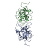
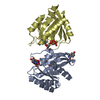
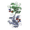
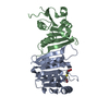
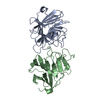
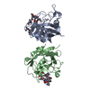
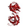
| Title | Crystal Structure of Murraya koenigii Miraculin-Like Protein at 2.2 A resolution at pH 4.6 |
|---|
 Components Components | TRYPSIN INHIBITOR |
|---|
 Keywords Keywords | HYDROLASE INHIBITOR / BETA-TREFOIL FOLD / KUNITZ SUPER-FAMILY |
|---|
| Function / homology |  Function and homology information Function and homology information
Soybean trypsin inhibitor (Kunitz) protease inhibitors family signature. / Proteinase inhibitor I3, Kunitz legume / Trypsin and protease inhibitor / Soybean trypsin inhibitor (Kunitz) family of protease inhibitors / Kunitz inhibitor STI-like superfamily / Trefoil (Acidic Fibroblast Growth Factor, subunit A) - #50 / Trefoil (Acidic Fibroblast Growth Factor, subunit A) / Trefoil / Mainly BetaSimilarity search - Domain/homology |
|---|
| Biological species |  MURRAYA KOENIGII (curry leaf) MURRAYA KOENIGII (curry leaf) |
|---|
| Method |  X-RAY DIFFRACTION / MOLECULAR REPLACEMENT / Resolution: 2.24 Å X-RAY DIFFRACTION / MOLECULAR REPLACEMENT / Resolution: 2.24 Å |
|---|
 Authors Authors | Selvakumar, P. / Sharma, N. / Tomar, P.P.S. / Kumar, P. / Sharma, A.K. |
|---|
 Citation Citation | Journal: Proteins / Year: 2014
Title: Structural Insights Into the Aggregation Behavior of Murraya Koenigii Miraculin-Like Protein Below Ph 7.5.
Authors: Selvakumar, P. / Sharma, N. / Tomar, P.P.S. / Kumar, P. / Sharma, A.K. |
|---|
| History | | Deposition | Nov 19, 2012 | Deposition site: PDBE / Processing site: PDBE |
|---|
| Revision 1.0 | Dec 4, 2013 | Provider: repository / Type: Initial release |
|---|
| Revision 1.1 | Apr 23, 2014 | Group: Database references |
|---|
| Revision 1.2 | Dec 20, 2023 | Group: Data collection / Database references ...Data collection / Database references / Other / Refinement description
Category: chem_comp_atom / chem_comp_bond ...chem_comp_atom / chem_comp_bond / database_2 / pdbx_database_status / pdbx_initial_refinement_model
Item: _database_2.pdbx_DOI / _database_2.pdbx_database_accession / _pdbx_database_status.status_code_sf |
|---|
| Revision 1.3 | Oct 16, 2024 | Group: Structure summary / Category: pdbx_entry_details / pdbx_modification_feature |
|---|
|
|---|
 Movie
Movie Controller

 Yorodumi
 Open data
 Basic information
 Structure visualization
 Downloads & links
 Other downloads
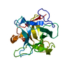


 PDBj
PDBj Assembly
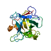
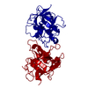
 Mass: 18.015 Da / Num. of mol.: 69 / Source method: isolated from a natural source / Formula: H2O
 Sample preparation
 Processing