 Movie
Movie Controller

[English] 日本語
 Yorodumi
- PDB-3x32: Crystal structure of the oxidized form of the solubilized domain ... -
+ Open data

- Basic information
| Entry | Database: PDB / ID: 3x32 | ||||||
|---|---|---|---|---|---|---|---|
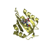
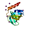
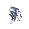
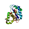
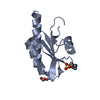
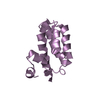
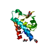
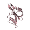
| Title | Crystal structure of the oxidized form of the solubilized domain of porcine cytochrome b5 in form 1 crystal | ||||||
 Components Components | Cytochrome b5 | ||||||
 Keywords Keywords | ELECTRON TRANSPORT / HEME | ||||||
| Function / homology |  Function and homology information Function and homology informationVitamin C (ascorbate) metabolism / Insertion of tail-anchored proteins into the endoplasmic reticulum membrane / heme binding / endoplasmic reticulum membrane / metal ion binding Similarity search - Function | ||||||
| Biological species |  | ||||||
| Method |  X-RAY DIFFRACTION / SYNCHROTRON / MOLECULAR REPLACEMENT / Resolution: 0.83 Å X-RAY DIFFRACTION / SYNCHROTRON / MOLECULAR REPLACEMENT / Resolution: 0.83 Å | ||||||
 Authors Authors | Hirano, Y. / Kimura, S. / Tamada, T. | ||||||
 Citation Citation | Journal: Acta Crystallogr.,Sect.D / Year: 2015 Title: High-resolution crystal structures of the solubilized domain of porcine cytochrome b5. Authors: Hirano, Y. / Kimura, S. / Tamada, T. | ||||||
| History |
|
- Structure visualization
| Structure viewer | Molecule: MolmilJmol/JSmol |
|---|
- Downloads & links
-Download
| PDBx/mmCIF format | 3x32.cif.gz | 80.1 KB | Display | PDBx/mmCIF format |
|---|---|---|---|---|
| PDB format | pdb3x32.ent.gz | 59.6 KB | Display | PDB format |
| PDBx/mmJSON format | 3x32.json.gz | Tree view | PDBx/mmJSON format | |
| Others |  Other downloads Other downloads |
-Validation report
| Arichive directory | https://data.pdbj.org/pub/pdb/validation_reports/x3/3x32ftp://data.pdbj.org/pub/pdb/validation_reports/x3/3x32 | HTTPS FTP |
|---|
-Related structure data
| Related structure data | 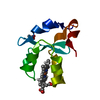 3x33C  3x34C 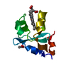 3x35C 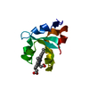 1cyoS C: citing same article ( S: Starting model for refinement |
|---|---|
| Similar structure data |

-Links
 PDBj
PDBj
- Assembly
| Deposited unit | 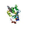
| ||||||||
|---|---|---|---|---|---|---|---|---|---|
| 1 |
| ||||||||
| Unit cell |
|
-Components
| #1: Protein | Mass: 10770.889 Da / Num. of mol.: 1 / Fragment: N-TERMINAL DOMAIN, UNP residues 1-94 Source method: isolated from a genetically manipulated source Source: (gene. exp.)  | ||||
|---|---|---|---|---|---|
| #2: Chemical | ChemComp-HEM /   Mass: 616.487 Da / Num. of mol.: 1 / Source method: obtained synthetically / Formula: C34H32FeN4O4 Mass: 616.487 Da / Num. of mol.: 1 / Source method: obtained synthetically / Formula: C34H32FeN4O4 | ||||
| #3: Chemical |   Mass: 40.078 Da / Num. of mol.: 2 / Source method: obtained synthetically / Formula: Ca Mass: 40.078 Da / Num. of mol.: 2 / Source method: obtained synthetically / Formula: Ca#4: Chemical | ChemComp-EPE / |   Mass: 238.305 Da / Num. of mol.: 1 / Source method: obtained synthetically / Formula: C8H18N2O4S / Comment: pH buffer*YM Mass: 238.305 Da / Num. of mol.: 1 / Source method: obtained synthetically / Formula: C8H18N2O4S / Comment: pH buffer*YM#5: Water | ChemComp-HOH / |  Mass: 18.015 Da / Num. of mol.: 214 / Source method: isolated from a natural source / Formula: H2O Mass: 18.015 Da / Num. of mol.: 214 / Source method: isolated from a natural source / Formula: H2O |
-Experimental details
-Experiment
| Experiment | Method: X-RAY DIFFRACTION / Number of used crystals: 1 |
|---|
- Sample preparation
| Crystal | Density Matthews: 1.8 Å3/Da / Density % sol: 31.55 % |
|---|---|
| Crystal grow | Temperature: 293 K / Method: vapor diffusion, hanging drop / pH: 7.5 Details: 24% PEG 1500, 4% 2-propanol, 0.1M calcium chloride, 0.1M HEPES-NaOH, pH 7.5, VAPOR DIFFUSION, HANGING DROP, temperature 293K |
-Data collection
| Diffraction | Mean temperature: 100 K |
|---|---|
| Diffraction source | Source: SYNCHROTRON / Site: Photon Factory  / Beamline: BL-17A / Wavelength: 0.91 Å / Beamline: BL-17A / Wavelength: 0.91 Å |
| Detector | Type: ADSC QUANTUM 270 / Detector: CCD / Date: Jun 9, 2014 |
| Radiation | Monochromator: Si 111 / Protocol: SINGLE WAVELENGTH / Monochromatic (M) / Laue (L): M / Scattering type: x-ray |
| Radiation wavelength | Wavelength: 0.91 Å / Relative weight: 1 |
| Reflection | Resolution: 0.83→50 Å / Num. all: 73045 / Num. obs: 72789 / % possible obs: 98.2 % / Observed criterion σ(F): 0 / Observed criterion σ(I): 0 / Redundancy: 15.5 % / Biso Wilson estimate: 6.2 Å2 / Rmerge(I) obs: 0.089 / Net I/σ(I): 59.8 |
| Reflection shell | Resolution: 0.83→0.84 Å / Redundancy: 10.8 % / Rmerge(I) obs: 0.374 / Mean I/σ(I) obs: 7.6 / % possible all: 82.3 |
- Processing
| Software |
| ||||||||||||||||||||
|---|---|---|---|---|---|---|---|---|---|---|---|---|---|---|---|---|---|---|---|---|---|
| Refinement | Method to determine structure: MOLECULAR REPLACEMENT Starting model: PDB ENTRY 1CYO Resolution: 0.83→50 Å / σ(F): 0 / Stereochemistry target values: Engh & Huber
| ||||||||||||||||||||
| Refinement step | Cycle: LAST / Resolution: 0.83→50 Å
| ||||||||||||||||||||
| Refine LS restraints |
|
