 Movie
Movie Controller

[English] 日本語
 Yorodumi
- PDB-3x2p: Neutron and X-ray joint refined structure of PcCel45A with cellop... -
+ Open data

- Basic information
| Entry | Database: PDB / ID: 3x2p | |||||||||
|---|---|---|---|---|---|---|---|---|---|---|
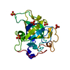
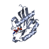
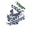
| Title | Neutron and X-ray joint refined structure of PcCel45A with cellopentaose at 298K. | |||||||||
 Components Components | Endoglucanase V-like protein | |||||||||
 Keywords Keywords | HYDROLASE | |||||||||
| Function / homology |  Function and homology information Function and homology informationExpansin/pollen allergen, DPBB domain / Expansin, family-45 endoglucanase-like domain profile. / EXPB1-like domain 1 / RlpA-like domain / RlpA-like domain superfamily / Barwin-like endoglucanases / Beta Barrel / Mainly Beta Similarity search - Domain/homology | |||||||||
| Biological species |  Phanerochaete chrysosporium (fungus) Phanerochaete chrysosporium (fungus) | |||||||||
| Method | NEUTRON DIFFRACTION /  X-RAY DIFFRACTION / SYNCHROTRON / Resolution: 1.518 Å X-RAY DIFFRACTION / SYNCHROTRON / Resolution: 1.518 Å | |||||||||
 Authors Authors | Nakamura, A. / Ishida, T. / Kusaka, K. / Yamada, T. / Tanaka, I. / Niimura, N. / Samejima, M. / Igarashi, K. | |||||||||
 Citation Citation | Journal: Sci Adv / Year: 2015 Title: "Newton's cradle" proton relay with amide-imidic acid tautomerization in inverting cellulase visualized by neutron crystallography. Authors: Nakamura, A. / Ishida, T. / Kusaka, K. / Yamada, T. / Fushinobu, S. / Tanaka, I. / Kaneko, S. / Ohta, K. / Tanaka, H. / Inaka, K. / Higuchi, Y. / Niimura, N. / Samejima, M. / Igarashi, K. | |||||||||
| History |
|
- Structure visualization
| Structure viewer | Molecule: MolmilJmol/JSmol |
|---|
- Downloads & links
-Download
| PDBx/mmCIF format | 3x2p.cif.gz | 124.3 KB | Display | PDBx/mmCIF format |
|---|---|---|---|---|
| PDB format | pdb3x2p.ent.gz | 99.7 KB | Display | PDB format |
| PDBx/mmJSON format | 3x2p.json.gz | Tree view | PDBx/mmJSON format | |
| Others |  Other downloads Other downloads |
-Validation report
| Arichive directory | https://data.pdbj.org/pub/pdb/validation_reports/x2/3x2pftp://data.pdbj.org/pub/pdb/validation_reports/x2/3x2p | HTTPS FTP |
|---|
-Related structure data
| Related structure data |  3x2gC  3x2hC  3x2iC  3x2jC  3x2kC  3x2lC  3x2mC  3x2nC  3x2oC 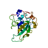 4zm7C C: citing same article ( |
|---|---|
| Similar structure data |


-Links
 PDBj
PDBj


- Assembly
| Deposited unit | 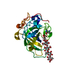
| ||||||||
|---|---|---|---|---|---|---|---|---|---|
| 1 |
| ||||||||
| Unit cell |
|
-Components
| #1: Protein | Mass: 18178.793 Da / Num. of mol.: 1 / Fragment: UNP residues 27-206 Source method: isolated from a genetically manipulated source Source: (gene. exp.) Phanerochaete chrysosporium (fungus) / Strain: K-3 / Gene: egv, PcCel45A / Plasmid: pPICZa / Production host: Pichia pastoris (fungus) / Strain (production host): KM71H / References: UniProt: B3Y002 |
|---|---|
| #2: Polysaccharide | beta-D-glucopyranose-(1-4)-beta-D-glucopyranose-(1-4)-beta-D-glucopyranose-(1-4)-beta-D- ...beta-D-glucopyranose-(1-4)-beta-D-glucopyranose-(1-4)-beta-D-glucopyranose-(1-4)-beta-D-glucopyranose-(1-4)-beta-D-glucopyranose / beta-cellopentaose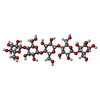  Source method: isolated from a genetically manipulated source Details: oligosaccharide / References: beta-cellopentaose |
| #3: Chemical | ChemComp-DOD /   Mass: 18.015 Da / Num. of mol.: 149 / Source method: isolated from a natural source / Formula: D2O Mass: 18.015 Da / Num. of mol.: 149 / Source method: isolated from a natural source / Formula: D2O |
| Has protein modification | Y |
-Experimental details
-Experiment
| Experiment |
|
|---|
- Sample preparation
| Crystal | Density Matthews: 2.43 Å3/Da / Density % sol: 49.31 % |
|---|---|
| Crystal grow | Temperature: 293 K / Method: vapor diffusion, sitting drop / pH: 8 Details: 60% 3-methyl-1,5-pentanediol, 50mM Tris-HCl, 2.5mM Cellopentaose, pH 8.0, VAPOR DIFFUSION, SITTING DROP, temperature 293K |
-Data collection
| Diffraction |
| ||||||||||||||||||||||||
|---|---|---|---|---|---|---|---|---|---|---|---|---|---|---|---|---|---|---|---|---|---|---|---|---|---|
| Diffraction source |
| ||||||||||||||||||||||||
| Detector |
| ||||||||||||||||||||||||
| Radiation |
| ||||||||||||||||||||||||
| Radiation wavelength |
| ||||||||||||||||||||||||
| Reflection | Entry-ID: 3X2P
| ||||||||||||||||||||||||
| Reflection shell |
|

- Processing
| Software |
| |||||||||||||||||||||||||||||||||||||||||||||||||||||||||||||||||||||||||||||||||||||||||||||||||||||||||||||||||||||||||||||||||||||
|---|---|---|---|---|---|---|---|---|---|---|---|---|---|---|---|---|---|---|---|---|---|---|---|---|---|---|---|---|---|---|---|---|---|---|---|---|---|---|---|---|---|---|---|---|---|---|---|---|---|---|---|---|---|---|---|---|---|---|---|---|---|---|---|---|---|---|---|---|---|---|---|---|---|---|---|---|---|---|---|---|---|---|---|---|---|---|---|---|---|---|---|---|---|---|---|---|---|---|---|---|---|---|---|---|---|---|---|---|---|---|---|---|---|---|---|---|---|---|---|---|---|---|---|---|---|---|---|---|---|---|---|---|---|---|
| Refinement | Shrinkage radii: 0.9 Å / VDW probe radii: 1.11 Å / Stereochemistry target values: ML / Solvent model: FLAT BULK SOLVENT MODEL
| |||||||||||||||||||||||||||||||||||||||||||||||||||||||||||||||||||||||||||||||||||||||||||||||||||||||||||||||||||||||||||||||||||||
| Refinement step | Cycle: LAST / Resolution: 1.518→19.685 Å
| |||||||||||||||||||||||||||||||||||||||||||||||||||||||||||||||||||||||||||||||||||||||||||||||||||||||||||||||||||||||||||||||||||||
| Refine LS restraints |
| |||||||||||||||||||||||||||||||||||||||||||||||||||||||||||||||||||||||||||||||||||||||||||||||||||||||||||||||||||||||||||||||||||||
| LS refinement shell |
|
