 Movie
Movie Controller


+ Open data

- Basic information
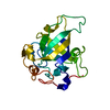
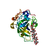
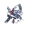
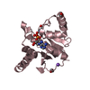
| Entry | Database: PDB / ID: 3x2n | ||||||
|---|---|---|---|---|---|---|---|
| Title | Proton relay pathway in inverting cellulase | ||||||
 Components Components | Endoglucanase V-like protein | ||||||
 Keywords Keywords | HYDROLASE | ||||||
| Function / homology | Expansin/pollen allergen, DPBB domain / Expansin, family-45 endoglucanase-like domain profile. / EXPB1-like domain 1 / RlpA-like domain / RlpA-like domain superfamily / Barwin-like endoglucanases / Beta Barrel / Mainly Beta / Endoglucanase V-like protein Function and homology information Function and homology information | ||||||
| Biological species |  Phanerochaete chrysosporium (fungus) Phanerochaete chrysosporium (fungus) | ||||||
| Method |  X-RAY DIFFRACTION / SYNCHROTRON / MIR / Resolution: 1.2 Å X-RAY DIFFRACTION / SYNCHROTRON / MIR / Resolution: 1.2 Å | ||||||
 Authors Authors | Nakamura, A. / Ishida, T. / Fushinobu, S. / Igarashi, K. / Samejima, M. | ||||||
 Citation Citation | Journal: Sci Adv / Year: 2015 Title: "Newton's cradle" proton relay with amide-imidic acid tautomerization in inverting cellulase visualized by neutron crystallography. Authors: Nakamura, A. / Ishida, T. / Kusaka, K. / Yamada, T. / Fushinobu, S. / Tanaka, I. / Kaneko, S. / Ohta, K. / Tanaka, H. / Inaka, K. / Higuchi, Y. / Niimura, N. / Samejima, M. / Igarashi, K. | ||||||
| History |
|
- Structure visualization
| Structure viewer | Molecule: MolmilJmol/JSmol |
|---|
- Downloads & links
-Download
| PDBx/mmCIF format | 3x2n.cif.gz | 91.9 KB | Display | PDBx/mmCIF format |
|---|---|---|---|---|
| PDB format | pdb3x2n.ent.gz | 68.9 KB | Display | PDB format |
| PDBx/mmJSON format | 3x2n.json.gz | Tree view | PDBx/mmJSON format | |
| Others |  Other downloads Other downloads |
-Validation report
| Arichive directory | https://data.pdbj.org/pub/pdb/validation_reports/x2/3x2nftp://data.pdbj.org/pub/pdb/validation_reports/x2/3x2n | HTTPS FTP |
|---|
-Related structure data
| Related structure data |  3x2gC 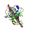 3x2hC 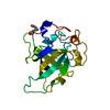 3x2iC  3x2jC  3x2kC  3x2lC  3x2mC 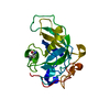 3x2oC  3x2pC 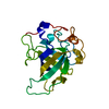 4zm7C C: citing same article ( |
|---|---|
| Similar structure data |


-Links
 PDBj
PDBj- Assembly
| Deposited unit | 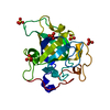
| ||||||||
|---|---|---|---|---|---|---|---|---|---|
| 1 |
| ||||||||
| Unit cell |
| ||||||||
| Components on special symmetry positions |
|
-Components
| #1: Protein | Mass: 18178.793 Da / Num. of mol.: 1 Source method: isolated from a genetically manipulated source Source: (gene. exp.) Phanerochaete chrysosporium (fungus) / Strain: K-3 / Gene: egv, PcCel45A / Plasmid: pPICZalpha / Production host: Pichia pastoris (fungus) / Strain (production host): KM71H / References: UniProt: B3Y002 | ||||
|---|---|---|---|---|---|
| #2: Chemical | ChemComp-SO4 /   Mass: 96.063 Da / Num. of mol.: 5 / Source method: obtained synthetically / Formula: SO4 Mass: 96.063 Da / Num. of mol.: 5 / Source method: obtained synthetically / Formula: SO4#3: Water | ChemComp-HOH / |  Mass: 18.015 Da / Num. of mol.: 338 / Source method: isolated from a natural source / Formula: H2O Mass: 18.015 Da / Num. of mol.: 338 / Source method: isolated from a natural source / Formula: H2OHas protein modification | Y | |
-Experimental details
-Experiment
| Experiment | Method: X-RAY DIFFRACTION / Number of used crystals: 1 |
|---|
- Sample preparation
| Crystal | Density Matthews: 1.97 Å3/Da / Density % sol: 37.55 % |
|---|---|
| Crystal grow | Temperature: 277 K / Method: vapor diffusion, sitting drop / pH: 8 Details: 3.0M Ammonium Sulphate, 20mM Tris-HCl, pH 8.0, VAPOR DIFFUSION, SITTING DROP, temperature 277K |
-Data collection
| Diffraction | Mean temperature: 95 K |
|---|---|
| Diffraction source | Source: SYNCHROTRON / Site: Photon Factory  / Beamline: BL-5A / Wavelength: 1 Å / Beamline: BL-5A / Wavelength: 1 Å |
| Detector | Type: ADSC QUANTUM 315r / Detector: CCD / Details: mirrors |
| Radiation | Protocol: SINGLE WAVELENGTH / Monochromatic (M) / Laue (L): M / Scattering type: x-ray |
| Radiation wavelength | Wavelength: 1 Å / Relative weight: 1 |
| Reflection | Resolution: 1.2→50 Å / Num. all: 44358 / Num. obs: 44001 / % possible obs: 99.2 % / Observed criterion σ(I): 663.6 / Redundancy: 3.6 % / Rsym value: 0.053 / Net I/σ(I): 34.92 |
| Reflection shell | Resolution: 1.2→1.22 Å / Redundancy: 3.5 % / Mean I/σ(I) obs: 15.96 / Num. unique all: 2204 / Rsym value: 0.085 / % possible all: 98.1 |
- Processing
| Software |
| ||||||||||||||||||||||||||||||||||||||||||||||||||||||||||||||||||||||||||||||||||||||||||||||||||||||||||||||||||||||||||||||||||||||||||||||||||||||||||||||||||||||||||||||||||||||
|---|---|---|---|---|---|---|---|---|---|---|---|---|---|---|---|---|---|---|---|---|---|---|---|---|---|---|---|---|---|---|---|---|---|---|---|---|---|---|---|---|---|---|---|---|---|---|---|---|---|---|---|---|---|---|---|---|---|---|---|---|---|---|---|---|---|---|---|---|---|---|---|---|---|---|---|---|---|---|---|---|---|---|---|---|---|---|---|---|---|---|---|---|---|---|---|---|---|---|---|---|---|---|---|---|---|---|---|---|---|---|---|---|---|---|---|---|---|---|---|---|---|---|---|---|---|---|---|---|---|---|---|---|---|---|---|---|---|---|---|---|---|---|---|---|---|---|---|---|---|---|---|---|---|---|---|---|---|---|---|---|---|---|---|---|---|---|---|---|---|---|---|---|---|---|---|---|---|---|---|---|---|---|---|
| Refinement | Method to determine structure: MIR / Resolution: 1.2→27.92 Å / Cor.coef. Fo:Fc: 0.977 / Cor.coef. Fo:Fc free: 0.968 / SU B: 0.785 / SU ML: 0.017 / Cross valid method: THROUGHOUT / ESU R: 0.033 / ESU R Free: 0.032 / Stereochemistry target values: MAXIMUM LIKELIHOOD
| ||||||||||||||||||||||||||||||||||||||||||||||||||||||||||||||||||||||||||||||||||||||||||||||||||||||||||||||||||||||||||||||||||||||||||||||||||||||||||||||||||||||||||||||||||||||
| Solvent computation | Ion probe radii: 0.8 Å / Shrinkage radii: 0.8 Å / VDW probe radii: 1.2 Å / Solvent model: MASK | ||||||||||||||||||||||||||||||||||||||||||||||||||||||||||||||||||||||||||||||||||||||||||||||||||||||||||||||||||||||||||||||||||||||||||||||||||||||||||||||||||||||||||||||||||||||
| Displacement parameters | Biso mean: 7.762 Å2
| ||||||||||||||||||||||||||||||||||||||||||||||||||||||||||||||||||||||||||||||||||||||||||||||||||||||||||||||||||||||||||||||||||||||||||||||||||||||||||||||||||||||||||||||||||||||
| Refinement step | Cycle: LAST / Resolution: 1.2→27.92 Å
| ||||||||||||||||||||||||||||||||||||||||||||||||||||||||||||||||||||||||||||||||||||||||||||||||||||||||||||||||||||||||||||||||||||||||||||||||||||||||||||||||||||||||||||||||||||||
| Refine LS restraints |
|