 Movie
Movie Controller

[English] 日本語
 Yorodumi
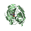
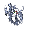
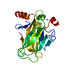
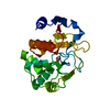
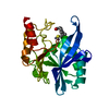
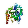
- PDB-2j92: 3C PROTEASE FROM TYPE A10(61) FOOT-AND-MOUTH DISEASE VIRUS - Crys... -
+ Open data

- Basic information
| Entry | Database: PDB / ID: 2j92 | ||||||
|---|---|---|---|---|---|---|---|
| Title | 3C PROTEASE FROM TYPE A10(61) FOOT-AND-MOUTH DISEASE VIRUS - Crystal packing mutant (K51Q) | ||||||
 Components Components | PICORNAIN 3C | ||||||
 Keywords Keywords | HYDROLASE / FOOT-AND- MOUTH DISEASE VIRUS / CHYMOTRYPSIN-LIKE CYSTEINE PROTEASE / THIOL PROTEASE / RNA REPLICATION | ||||||
| Function / homology |  Function and homology information Function and homology informationL-peptidase / symbiont-mediated perturbation of host chromatin organization / picornain 3C / T=pseudo3 icosahedral viral capsid / host cell cytoplasmic vesicle membrane / ribonucleoside triphosphate phosphatase activity / nucleoside-triphosphate phosphatase / channel activity / monoatomic ion transmembrane transport / clathrin-dependent endocytosis of virus by host cell ...L-peptidase / symbiont-mediated perturbation of host chromatin organization / picornain 3C / T=pseudo3 icosahedral viral capsid / host cell cytoplasmic vesicle membrane / ribonucleoside triphosphate phosphatase activity / nucleoside-triphosphate phosphatase / channel activity / monoatomic ion transmembrane transport / clathrin-dependent endocytosis of virus by host cell / RNA helicase activity / viral protein processing / host cell endoplasmic reticulum membrane / symbiont-mediated activation of host autophagy / RNA-directed RNA polymerase / cysteine-type endopeptidase activity / viral RNA genome replication / RNA-directed RNA polymerase activity / virion attachment to host cell / host cell nucleus / DNA-templated transcription / structural molecule activity / proteolysis / RNA binding / ATP binding Similarity search - Function | ||||||
| Biological species |   FOOT-AND-MOUTH DISEASE VIRUS FOOT-AND-MOUTH DISEASE VIRUS | ||||||
| Method |  X-RAY DIFFRACTION / SYNCHROTRON / MOLECULAR REPLACEMENT / Resolution: 2.2 Å X-RAY DIFFRACTION / SYNCHROTRON / MOLECULAR REPLACEMENT / Resolution: 2.2 Å | ||||||
 Authors Authors | Sweeney, T.R. / Birtley, J.R. / Leatherbarrow, R.J. / Curry, S. | ||||||
 Citation Citation | Journal: J.Virol. / Year: 2007 Title: Structural and Mutagenic Analysis of Foot-and-Mouth Disease Virus 3C Protease Reveals the Role of the {Beta}-Ribbon in Proteolysis. Authors: Sweeney, T.R. / Roque-Rosell, N. / Birtley, J.R. / Leatherbarrow, R.J. / Curry, S. #1: Journal: J.Biol.Chem. / Year: 2005Title: Crystal Structure of Foot-and-Mouth Disease Virus 3C Protease: New Insights Into Catalytic Mechanism and Cleavage Specificity Authors: Birtley, J.R. / Knox, S.R. / Jaulent, A.M. / Brick, P. / Leatherbarrow, R.J. / Curry, S. | ||||||
| History |
| ||||||
| Remark 700 | SHEET DETERMINATION METHOD: DSSP THE SHEETS PRESENTED AS "AB" IN EACH CHAIN ON SHEET RECORDS BELOW ... SHEET DETERMINATION METHOD: DSSP THE SHEETS PRESENTED AS "AB" IN EACH CHAIN ON SHEET RECORDS BELOW IS ACTUALLY AN 12-STRANDED BARREL THIS IS REPRESENTED BY A 13-STRANDED SHEET IN WHICH THE FIRST AND LAST STRANDS ARE IDENTICAL. |
- Structure visualization
| Structure viewer | Molecule: MolmilJmol/JSmol |
|---|
- Downloads & links
-Download
| PDBx/mmCIF format | 2j92.cif.gz | 85.9 KB | Display | PDBx/mmCIF format |
|---|---|---|---|---|
| PDB format | pdb2j92.ent.gz | 64 KB | Display | PDB format |
| PDBx/mmJSON format | 2j92.json.gz | Tree view | PDBx/mmJSON format | |
| Others |  Other downloads Other downloads |
-Validation report
| Arichive directory | https://data.pdbj.org/pub/pdb/validation_reports/j9/2j92ftp://data.pdbj.org/pub/pdb/validation_reports/j9/2j92 | HTTPS FTP |
|---|
-Related structure data
| Related structure data | 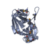 2bhgS S: Starting model for refinement |
|---|---|
| Similar structure data |


-Links
 PDBj
PDBj

- Assembly
| Deposited unit | 
| ||||||||
|---|---|---|---|---|---|---|---|---|---|
| 1 | 
| ||||||||
| 2 | 
| ||||||||
| Unit cell |
| ||||||||
| Noncrystallographic symmetry (NCS) | NCS oper: (Code: given Matrix: (-0.9227, 0.3129, 0.2253), Vector: |
-Components
| #1: Protein | Mass: 22518.006 Da / Num. of mol.: 2 / Mutation: YES Source method: isolated from a genetically manipulated source Details: K51Q - TO DISRUPT ORIGINAL CRYSTAL PACKING C95K - TO AVOID AGGREGATION C142S - TO AVOID AGGREGATION C163A - TO REMOVE ACITVE-STE NUCLEOPHILE. Source: (gene. exp.) FOOT-AND-MOUTH DISEASE VIRUS (STRAIN A10-61)Production host:  #2: Water | ChemComp-HOH / |  Mass: 18.015 Da / Num. of mol.: 109 / Source method: isolated from a natural source / Formula: H2O Mass: 18.015 Da / Num. of mol.: 109 / Source method: isolated from a natural source / Formula: H2OCompound details | ENGINEERED RESIDUE IN CHAIN A, LYS 1700 TO GLN ENGINEERED RESIDUE IN CHAIN A, CYS 1744 TO LYS ...ENGINEERED | Sequence details | THE CONFLICT ASN1671 (ILE22) ARISES PROBABLY BECAUSE OF A SEQUENCING ERROR IN THE P03306 ENTRY. ...THE CONFLICT ASN1671 (ILE22) ARISES PROBABLY BECAUSE OF A SEQUENCING | |
|---|
-Experimental details
-Experiment
| Experiment | Method: X-RAY DIFFRACTION / Number of used crystals: 1 |
|---|
- Sample preparation
| Crystal | Density Matthews: 2.4 Å3/Da / Density % sol: 48.36 % |
|---|---|
| Crystal grow | pH: 7 / Details: SEE PAPER, pH 7.00 |
-Data collection
| Diffraction | Mean temperature: 100 K |
|---|---|
| Diffraction source | Source: SYNCHROTRON / Site: SRS  / Beamline: PX10.1 / Wavelength: 1.488 / Beamline: PX10.1 / Wavelength: 1.488 |
| Detector | Type: MARRESEARCH / Detector: CCD / Date: Dec 15, 2005 Details: RH COATED COLLIMATING MIRROR, A DOUBLE CRYSTAL SI(III) MONOCHROMATOR WITH HORIZONTAL SAGGITAL FOCUSING SYSTEM, AND FINALLY A SECOND RH COATED MIRROR FOR VERTICAL FOCUSING. |
| Radiation | Monochromator: DOUBLE CRYSTAL SI(III) MONOCHROMATOR / Protocol: SINGLE WAVELENGTH / Monochromatic (M) / Laue (L): M / Scattering type: x-ray |
| Radiation wavelength | Wavelength: 1.488 Å / Relative weight: 1 |
| Reflection | Resolution: 2.2→23.6 Å / Num. obs: 20590 / % possible obs: 99.7 % / Observed criterion σ(I): 3 / Redundancy: 3.5 % / Biso Wilson estimate: 26.9 Å2 / Rmerge(I) obs: 0.08 / Net I/σ(I): 11.6 |
| Reflection shell | Resolution: 2.2→2.32 Å / Redundancy: 3.5 % / Rmerge(I) obs: 0.34 / Mean I/σ(I) obs: 3.8 / % possible all: 99.5 |
- Processing
| Software |
| ||||||||||||||||||||||||||||||||||||||||||||||||||||||||||||
|---|---|---|---|---|---|---|---|---|---|---|---|---|---|---|---|---|---|---|---|---|---|---|---|---|---|---|---|---|---|---|---|---|---|---|---|---|---|---|---|---|---|---|---|---|---|---|---|---|---|---|---|---|---|---|---|---|---|---|---|---|---|
| Refinement | Method to determine structure: MOLECULAR REPLACEMENT Starting model: PDB ENTRY 2BHG Resolution: 2.2→23.59 Å / Rfactor Rfree error: 0.008 / Data cutoff high absF: 1483613.08 / Isotropic thermal model: RESTRAINED / Cross valid method: THROUGHOUT / σ(F): 0 / Stereochemistry target values: MLF
| ||||||||||||||||||||||||||||||||||||||||||||||||||||||||||||
| Solvent computation | Solvent model: FLAT MODEL / Bsol: 31.3037 Å2 / ksol: 0.329538 e/Å3 | ||||||||||||||||||||||||||||||||||||||||||||||||||||||||||||
| Displacement parameters | Biso mean: 34.7 Å2
| ||||||||||||||||||||||||||||||||||||||||||||||||||||||||||||
| Refine analyze |
| ||||||||||||||||||||||||||||||||||||||||||||||||||||||||||||
| Refinement step | Cycle: LAST / Resolution: 2.2→23.59 Å
| ||||||||||||||||||||||||||||||||||||||||||||||||||||||||||||
| Refine LS restraints |
| ||||||||||||||||||||||||||||||||||||||||||||||||||||||||||||
| LS refinement shell | Resolution: 2.2→2.34 Å / Rfactor Rfree error: 0.031 / Total num. of bins used: 6
| ||||||||||||||||||||||||||||||||||||||||||||||||||||||||||||
| Xplor file |
|
