 Movie
Movie Controller


+ Open data

- Basic information
| Entry | Database: PDB / ID: 3wql | ||||||
|---|---|---|---|---|---|---|---|
| Title | Crystal structure of Rv3378c with Mg2+ and PPi | ||||||
 Components Components | Diterpene synthase | ||||||
 Keywords Keywords | HYDROLASE / phosphatase / diterpene synthase | ||||||
| Function / homology |  Function and homology information Function and homology informationadenosine tuberculosinyltransferase / (13S)-vitexifolin A synthase activity / tuberculosinol biosynthetic process / diterpenoid biosynthetic process / transferase activity, transferring alkyl or aryl (other than methyl) groups / phosphatase activity Similarity search - Function | ||||||
| Biological species |   Mycobacterium tuberculosis (bacteria) Mycobacterium tuberculosis (bacteria) | ||||||
| Method |  X-RAY DIFFRACTION / SYNCHROTRON / MOLECULAR REPLACEMENT / Resolution: 2.1 Å X-RAY DIFFRACTION / SYNCHROTRON / MOLECULAR REPLACEMENT / Resolution: 2.1 Å | ||||||
 Authors Authors | Chan, H.C. / Feng, X. / Ko, T.P. / Huang, C.H. / Hu, Y. / Zheng, Y. / Bogue, S. / Nakano, C. / Hoshino, T. / Zhang, L. ...Chan, H.C. / Feng, X. / Ko, T.P. / Huang, C.H. / Hu, Y. / Zheng, Y. / Bogue, S. / Nakano, C. / Hoshino, T. / Zhang, L. / Lv, P. / Liu, W. / Crick, D.C. / Liang, P.H. / Wang, A.H. / Oldfield, E. / Guo, R.T. | ||||||
 Citation Citation | Journal: J.Am.Chem.Soc. / Year: 2014 Title: Structure and inhibition of tuberculosinol synthase and decaprenyl diphosphate synthase from Mycobacterium tuberculosis. Authors: Chan, H.C. / Feng, X. / Ko, T.P. / Huang, C.H. / Hu, Y. / Zheng, Y. / Bogue, S. / Nakano, C. / Hoshino, T. / Zhang, L. / Lv, P. / Liu, W. / Crick, D.C. / Liang, P.H. / Wang, A.H. / Oldfield, E. / Guo, R.T. | ||||||
| History |
|
- Structure visualization
| Structure viewer | Molecule: MolmilJmol/JSmol |
|---|
- Downloads & links
-Download
| PDBx/mmCIF format | 3wql.cif.gz | 257 KB | Display | PDBx/mmCIF format |
|---|---|---|---|---|
| PDB format | pdb3wql.ent.gz | 207.8 KB | Display | PDB format |
| PDBx/mmJSON format | 3wql.json.gz | Tree view | PDBx/mmJSON format | |
| Others |  Other downloads Other downloads |
-Validation report
| Arichive directory | https://data.pdbj.org/pub/pdb/validation_reports/wq/3wqlftp://data.pdbj.org/pub/pdb/validation_reports/wq/3wql | HTTPS FTP |
|---|
-Related structure data
| Related structure data |  3wqkSC  3wqmC  3wqnC  4kt8C  4oncC S: Starting model for refinement C: citing same article ( |
|---|---|
| Similar structure data |


-Links
 PDBj
PDBj- Assembly
| Deposited unit | 
| ||||||||
|---|---|---|---|---|---|---|---|---|---|
| 1 | 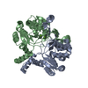
| ||||||||
| 2 | 
| ||||||||
| Unit cell |
| ||||||||
| Components on special symmetry positions |
|
-Components
| #1: Protein | Mass: 34071.484 Da / Num. of mol.: 4 Source method: isolated from a genetically manipulated source Source: (gene. exp.) Mycobacterium tuberculosis (bacteria) / Strain: H37Rv / Gene: Rv3378c, MT3488 / Production host: References: UniProt: O50407, UniProt: P9WJ61*PLUS, EC: 3.1.7.9, EC: 3.1.7.8 #2: Chemical | ChemComp-MG /   Mass: 24.305 Da / Num. of mol.: 8 / Source method: obtained synthetically / Formula: Mg Mass: 24.305 Da / Num. of mol.: 8 / Source method: obtained synthetically / Formula: Mg#3: Water | ChemComp-HOH / |  Mass: 18.015 Da / Num. of mol.: 1018 / Source method: isolated from a natural source / Formula: H2O Mass: 18.015 Da / Num. of mol.: 1018 / Source method: isolated from a natural source / Formula: H2O |
|---|
-Experimental details
-Experiment
| Experiment | Method: X-RAY DIFFRACTION |
|---|
- Sample preparation
| Crystal | Density Matthews: 2.47 Å3/Da / Density % sol: 50.26 % |
|---|
-Data collection
| Diffraction | Mean temperature: 100 K |
|---|---|
| Diffraction source | Source: SYNCHROTRON / Site: NSRRC  / Beamline: BL13C1 / Wavelength: 1 Å / Beamline: BL13C1 / Wavelength: 1 Å |
| Detector | Type: ADSC QUANTUM 315r / Detector: CCD / Date: Nov 6, 2011 |
| Radiation | Protocol: SINGLE WAVELENGTH / Monochromatic (M) / Laue (L): M / Scattering type: x-ray |
| Radiation wavelength | Wavelength: 1 Å / Relative weight: 1 |
| Reflection | Resolution: 2.1→25 Å / Num. obs: 77301 / % possible obs: 98.1 % / Redundancy: 3.9 % / Rmerge(I) obs: 0.065 |
| Reflection shell | Resolution: 2.1→2.2 Å / Redundancy: 2.7 % / Rmerge(I) obs: 0.416 / Mean I/σ(I) obs: 2.3 / % possible all: 85.6 |
- Processing
| Software |
| ||||||||||||||||||
|---|---|---|---|---|---|---|---|---|---|---|---|---|---|---|---|---|---|---|---|
| Refinement | Method to determine structure: MOLECULAR REPLACEMENT Starting model: 3WQK Resolution: 2.1→25 Å / σ(F): 2 / Stereochemistry target values: Engh & Huber
| ||||||||||||||||||
| Refinement step | Cycle: LAST / Resolution: 2.1→25 Å
| ||||||||||||||||||
| LS refinement shell | Resolution: 2.1→2.2 Å /
|
